Daniele Veritti, Valentina Sarao, Marco Gonfiantini, Leopoldo Rubinato, Paolo Lanzetta
{"title":"Faricimab in Neovascular AMD Complicated by Pigment Epithelium Detachment: An AI-Assisted Evaluation of Early Morphological Changes.","authors":"Daniele Veritti, Valentina Sarao, Marco Gonfiantini, Leopoldo Rubinato, Paolo Lanzetta","doi":"10.1007/s40123-024-01005-x","DOIUrl":null,"url":null,"abstract":"<p><strong>Introduction: </strong>This study investigates the early temporal changes in pigment epithelial detachment (PED) morphology following treatment with faricimab in patients with neovascular age-related macular degeneration (nAMD). Utilizing an artificial intelligence (AI)-assisted approach, we provide a detailed quantification and characterization of the dynamics of these morphological changes.</p><p><strong>Methods: </strong>A prospective observational study was conducted on 22 eyes from 22 treatment-naïve patients with nAMD-associated PED (presenting either type 1 or type 3 macular neovascularization). Participants were administered intravitreal faricimab (6 mg) at baseline and at days 30, 60, and 90. Comprehensive ophthalmic evaluations and spectral-domain optical coherence tomography (SD-OCT) imaging were conducted at baseline and at seven additional follow-up visits on days 1, 7, 14, 30, 60, 90, and 120. An AI-based automated segmentation algorithm was utilized to precisely quantify changes in PED volume, alongside intraretinal (IRF) and subretinal fluid (SRF) volumes, at each time point.</p><p><strong>Results: </strong>Treatment with faricimab resulted in a significant reduction in mean PED volume, with an average decrease of 12% at day 1, 29% at day 7, 51% at day 14, 68% at day 30, 72% at day 60, 79% at day 90, and 84% at day 120 (p < 0.0001 for all time points). Similarly rapid and marked reductions were noted in both mean IRF (23.5% at day 1, 90.7% at day 14) and SRF (14.4% at day 1, 91.2% at day 14) volumes. The study also showed a statistically significant improvement in best-corrected visual acuity (BCVA) over the follow-up period, correlating with the reduction in PED volume.</p><p><strong>Conclusion: </strong>Faricimab demonstrates early and significant efficacy in improving PED architecture in patients with nAMD. The rapid morphological improvements observed in this study suggest faricimab may represent a valid therapeutic option for PEDs associated with nAMD.</p>","PeriodicalId":19623,"journal":{"name":"Ophthalmology and Therapy","volume":null,"pages":null},"PeriodicalIF":2.6000,"publicationDate":"2024-10-01","publicationTypes":"Journal Article","fieldsOfStudy":null,"isOpenAccess":false,"openAccessPdf":"https://www.ncbi.nlm.nih.gov/pmc/articles/PMC11408430/pdf/","citationCount":"0","resultStr":null,"platform":"Semanticscholar","paperid":null,"PeriodicalName":"Ophthalmology and Therapy","FirstCategoryId":"3","ListUrlMain":"https://doi.org/10.1007/s40123-024-01005-x","RegionNum":3,"RegionCategory":"医学","ArticlePicture":[],"TitleCN":null,"AbstractTextCN":null,"PMCID":null,"EPubDate":"2024/8/9 0:00:00","PubModel":"Epub","JCR":"Q2","JCRName":"OPHTHALMOLOGY","Score":null,"Total":0}
引用次数: 0
Abstract
Introduction: This study investigates the early temporal changes in pigment epithelial detachment (PED) morphology following treatment with faricimab in patients with neovascular age-related macular degeneration (nAMD). Utilizing an artificial intelligence (AI)-assisted approach, we provide a detailed quantification and characterization of the dynamics of these morphological changes.
Methods: A prospective observational study was conducted on 22 eyes from 22 treatment-naïve patients with nAMD-associated PED (presenting either type 1 or type 3 macular neovascularization). Participants were administered intravitreal faricimab (6 mg) at baseline and at days 30, 60, and 90. Comprehensive ophthalmic evaluations and spectral-domain optical coherence tomography (SD-OCT) imaging were conducted at baseline and at seven additional follow-up visits on days 1, 7, 14, 30, 60, 90, and 120. An AI-based automated segmentation algorithm was utilized to precisely quantify changes in PED volume, alongside intraretinal (IRF) and subretinal fluid (SRF) volumes, at each time point.
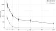
Results: Treatment with faricimab resulted in a significant reduction in mean PED volume, with an average decrease of 12% at day 1, 29% at day 7, 51% at day 14, 68% at day 30, 72% at day 60, 79% at day 90, and 84% at day 120 (p < 0.0001 for all time points). Similarly rapid and marked reductions were noted in both mean IRF (23.5% at day 1, 90.7% at day 14) and SRF (14.4% at day 1, 91.2% at day 14) volumes. The study also showed a statistically significant improvement in best-corrected visual acuity (BCVA) over the follow-up period, correlating with the reduction in PED volume.
Conclusion: Faricimab demonstrates early and significant efficacy in improving PED architecture in patients with nAMD. The rapid morphological improvements observed in this study suggest faricimab may represent a valid therapeutic option for PEDs associated with nAMD.
期刊介绍:
Aims and Scope
Ophthalmology and Therapy is an international, open access, peer-reviewed (single-blind), and rapid publication journal. The scope of the journal is broad and will consider all scientifically sound research from preclinical, clinical (all phases), observational, real-world, and health outcomes research around the use of ophthalmological therapies, devices, and surgical techniques.
The journal is of interest to a broad audience of pharmaceutical and healthcare professionals and publishes original research, reviews, case reports/series, trial protocols and short communications such as commentaries and editorials. Ophthalmology and Therapy will consider all scientifically sound research be it positive, confirmatory or negative data. Submissions are welcomed whether they relate to an international and/or a country-specific audience, something that is crucially important when researchers are trying to target more specific patient populations. This inclusive approach allows the journal to assist in the dissemination of quality research, which may be considered of insufficient interest by other journals.
Rapid Publication
The journal’s publication timelines aim for a rapid peer review of 2 weeks. If an article is accepted it will be published 3–4 weeks from acceptance. The rapid timelines are achieved through the combination of a dedicated in-house editorial team, who manage article workflow, and an extensive Editorial and Advisory Board who assist with peer review. This allows the journal to support the rapid dissemination of research, whilst still providing robust peer review. Combined with the journal’s open access model this allows for the rapid, efficient communication of the latest research and reviews, fostering the advancement of ophthalmic therapies.
Open Access
All articles published by Ophthalmology and Therapy are open access.
Personal Service
The journal’s dedicated in-house editorial team offer a personal “concierge service” meaning authors will always have an editorial contact able to update them on the status of their manuscript. The editorial team check all manuscripts to ensure that articles conform to the most recent COPE, GPP and ICMJE publishing guidelines. This supports the publication of ethically sound and transparent research.
Digital Features and Plain Language Summaries
Ophthalmology and Therapy offers a range of additional features designed to increase the visibility, readership and educational value of the journal’s content. Each article is accompanied by key summary points, giving a time-efficient overview of the content to a wide readership. Articles may be accompanied by plain language summaries to assist readers who have some knowledge of, but not in-depth expertise in, the area to understand the scientific content and overall implications of the article. The journal also provides the option to include various types of digital features including animated abstracts, video abstracts, slide decks, audio slides, instructional videos, infographics, podcasts and animations. All additional features are peer reviewed to the same high standard as the article itself. If you consider that your paper would benefit from the inclusion of a digital feature, please let us know. Our editorial team are able to create high-quality slide decks and infographics in-house, and video abstracts through our partner Research Square, and would be happy to assist in any way we can. For further information about digital features, please contact the journal editor (see ‘Contact the Journal’ for email address), and see the ‘Guidelines for digital features and plain language summaries’ document under ‘Submission guidelines’.
For examples of digital features please visit our showcase page https://springerhealthcare.com/expertise/publishing-digital-features/
Publication Fees
Upon acceptance of an article, authors will be required to pay the mandatory Rapid Service Fee of €5250/$6000/£4300. The journal will consider fee discounts and waivers for developing countries and this is decided on a case by case basis.
Peer Review Process
Upon submission, manuscripts are assessed by the editorial team to ensure they fit within the aims and scope of the journal and are also checked for plagiarism. All suitable submissions are then subject to a comprehensive single-blind peer review. Reviewers are selected based on their relevant expertise and publication history in the subject area. The journal has an extensive pool of editorial and advisory board members who have been selected to assist with peer review based on the afore-mentioned criteria.
At least two extensive reviews are required to make the editorial decision, with the exception of some article types such as Commentaries, Editorials, and Letters which are generally reviewed by one member of the Editorial Board. Where reviewer recommendations are conflicted, the editorial board will be contacted for further advice and a presiding decision. Manuscripts are then either accepted, rejected or authors are required to make major or minor revisions (both reviewer comments and editorial comments may need to be addressed). Once a revised manuscript is re-submitted, it is assessed along with the responses to reviewer comments and if it has been adequately revised it will be accepted for publication. Accepted manuscripts are then copyedited and typeset by the production team before online publication. Appeals against decisions following peer review are considered on a case-by-case basis and should be sent to the journal editor.
Preprints
We encourage posting of preprints of primary research manuscripts on preprint servers, authors’ or institutional websites, and open communications between researchers whether on community preprint servers or preprint commenting platforms. Posting of preprints is not considered prior publication and will not jeopardize consideration in our journals. Authors should disclose details of preprint posting during the submission process or at any other point during consideration in one of our journals. Once the manuscript is published, it is the author’s responsibility to ensure that the preprint record is updated with a publication reference, including the DOI and a URL link to the published version of the article on the journal website.
Please follow the link for further information on preprint sharing:
https://www.springer.com/gp/authors-editors/journal-author/journal-author-helpdesk/submission/1302#c16721550
Copyright
Ophthalmology and Therapy''s content is published open access under the Creative Commons Attribution-Noncommercial License, which allows users to read, copy, distribute, and make derivative works for non-commercial purposes from the material, as long as the author of the original work is cited. The author assigns the exclusive right to any commercial use of the article to Springer. For more information about the Creative Commons Attribution-Noncommercial License, click here: http://creativecommons.org/licenses/by-nc/4.0.
Contact
For more information about the journal, including pre-submission enquiries, please contact christopher.vautrinot@springer.com.

 求助内容:
求助内容: 应助结果提醒方式:


