Transarterial Embolization Enhances Programmed Cell Death Ligand 1 Expression and Influences CD8+T Lymphocytes Cytotoxicity in an Orthotopic Hepatocellular Carcinoma Rat Model.
Shen Zhang, Lin Xu, Jia-Qing Li, Ming-Zhan Du, Yu Yin, Bin-Yan Zhong, Han-Si Liang, Wan-Ci Li, Cai-Fang Ni, Xiao-Li Zhu
{"title":"Transarterial Embolization Enhances Programmed Cell Death Ligand 1 Expression and Influences CD8<sup>+</sup>T Lymphocytes Cytotoxicity in an Orthotopic Hepatocellular Carcinoma Rat Model.","authors":"Shen Zhang, Lin Xu, Jia-Qing Li, Ming-Zhan Du, Yu Yin, Bin-Yan Zhong, Han-Si Liang, Wan-Ci Li, Cai-Fang Ni, Xiao-Li Zhu","doi":"10.1007/s00270-024-03813-x","DOIUrl":null,"url":null,"abstract":"<p><strong>Purpose: </strong>To investigate the influence of transarterial embolization (TAE) on programmed cell death-ligand 1(PD-L1) expression and CD8<sup>+</sup>T tumour infiltrative lymphocyte cytotoxicity in the Sprague-Dawley (SD) rat model of hepatocellular carcinoma (HCC).</p><p><strong>Materials and methods: </strong>An orthotopic HCC model was established in twenty SD rats treated with TAE (lipiodol, n = 10) or sham (normal saline, n = 10) using homologous N1S1 hepatoma cells. Rats were euthanized 1 week after embolization. Flow cytometry was used to assess the proportion of CD4<sup>+</sup>T, CD8<sup>+</sup>T and programmed cell death-1<sup>+</sup>(PD-1<sup>+</sup>) CD8<sup>+</sup>T lymphocytes in the spleens and tumours. Distribution of CD8<sup>+</sup>T, granzyme-B<sup>+</sup>CD8<sup>+</sup>T lymphocytes and PD-L1<sup>+</sup> cells was assessed by immunohistochemistry (IHC) or multiplex IHC. p value < 0.05 was considered statistically significant.</p><p><strong>Results: </strong>The CD4/CD8 ratio and PD-1<sup>+</sup>CD8<sup>+</sup> T lymphocytes exhibited higher values in TAE-treated tumours compared to sham-treated tumours (p = 0.021 and p = 0.071, respectively). Conversely, the number of CD8<sup>+</sup>T lymphocytes was decreased in TAE-treated tumours (p = 0.043), especially in the central region (p = 0.045). However, more CD8<sup>+</sup>T lymphocytes were found infiltrating the marginal region than central region in TAE-treated tumours (p = 0.046). The proportion of granzyme-B<sup>+</sup>CD8<sup>+</sup>T lymphocytes and the PD-L1 positive areas was elevated in tumours that treated with TAE (p all < 0.05). There was a negative correlation between PD-L1 expression and the number of infiltration of CD8<sup>+</sup> T lymphocytes (p = 0.036).</p><p><strong>Conclusions: </strong>Immune cells are distributed unevenly in the tumours after TAE. The intrinsic induction state of the tumour after embolization may be insufficient to elicit a maximal response to PD-1/PD-L1 inhibitors.</p>","PeriodicalId":9591,"journal":{"name":"CardioVascular and Interventional Radiology","volume":null,"pages":null},"PeriodicalIF":2.8000,"publicationDate":"2024-08-05","publicationTypes":"Journal Article","fieldsOfStudy":null,"isOpenAccess":false,"openAccessPdf":"","citationCount":"0","resultStr":null,"platform":"Semanticscholar","paperid":null,"PeriodicalName":"CardioVascular and Interventional Radiology","FirstCategoryId":"3","ListUrlMain":"https://doi.org/10.1007/s00270-024-03813-x","RegionNum":3,"RegionCategory":"医学","ArticlePicture":[],"TitleCN":null,"AbstractTextCN":null,"PMCID":null,"EPubDate":"","PubModel":"","JCR":"Q2","JCRName":"CARDIAC & CARDIOVASCULAR SYSTEMS","Score":null,"Total":0}
引用次数: 0
Abstract
Purpose: To investigate the influence of transarterial embolization (TAE) on programmed cell death-ligand 1(PD-L1) expression and CD8+T tumour infiltrative lymphocyte cytotoxicity in the Sprague-Dawley (SD) rat model of hepatocellular carcinoma (HCC).
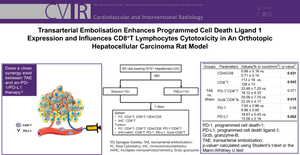
Materials and methods: An orthotopic HCC model was established in twenty SD rats treated with TAE (lipiodol, n = 10) or sham (normal saline, n = 10) using homologous N1S1 hepatoma cells. Rats were euthanized 1 week after embolization. Flow cytometry was used to assess the proportion of CD4+T, CD8+T and programmed cell death-1+(PD-1+) CD8+T lymphocytes in the spleens and tumours. Distribution of CD8+T, granzyme-B+CD8+T lymphocytes and PD-L1+ cells was assessed by immunohistochemistry (IHC) or multiplex IHC. p value < 0.05 was considered statistically significant.
Results: The CD4/CD8 ratio and PD-1+CD8+ T lymphocytes exhibited higher values in TAE-treated tumours compared to sham-treated tumours (p = 0.021 and p = 0.071, respectively). Conversely, the number of CD8+T lymphocytes was decreased in TAE-treated tumours (p = 0.043), especially in the central region (p = 0.045). However, more CD8+T lymphocytes were found infiltrating the marginal region than central region in TAE-treated tumours (p = 0.046). The proportion of granzyme-B+CD8+T lymphocytes and the PD-L1 positive areas was elevated in tumours that treated with TAE (p all < 0.05). There was a negative correlation between PD-L1 expression and the number of infiltration of CD8+ T lymphocytes (p = 0.036).
Conclusions: Immune cells are distributed unevenly in the tumours after TAE. The intrinsic induction state of the tumour after embolization may be insufficient to elicit a maximal response to PD-1/PD-L1 inhibitors.
期刊介绍:
CardioVascular and Interventional Radiology (CVIR) is the official journal of the Cardiovascular and Interventional Radiological Society of Europe, and is also the official organ of a number of additional distinguished national and international interventional radiological societies. CVIR publishes double blinded peer-reviewed original research work including clinical and laboratory investigations, technical notes, case reports, works in progress, and letters to the editor, as well as review articles, pictorial essays, editorials, and special invited submissions in the field of vascular and interventional radiology. Beside the communication of the latest research results in this field, it is also the aim of CVIR to support continuous medical education. Articles that are accepted for publication are done so with the understanding that they, or their substantive contents, have not been and will not be submitted to any other publication.
 求助内容:
求助内容: 应助结果提醒方式:


