{"title":"Convexity dural arteriovenous fistula with Sylvian-Labbé collateral pattern: A case report.","authors":"Phyo Wint Shwe Yee, Tatebayashi Kotaro, Uchida Kazutaka, Yoshimura Shinichi","doi":"10.7461/jcen.2024.E2024.05.001","DOIUrl":null,"url":null,"abstract":"<p><p>Convexity dural arteriovenous fistula (dAVF) is associated with high-grade dAVF and is usually presented with aggressive clinical presentation. Precise diagnosis and understanding the pathogenesis are important to achieving successful treatment without complications. We report a case of dAVF with Sylvian-Labbé collateral pattern, concerning embryological development that was thought to be involved in the vascular architecture and pathogenesis of dural AVF. Thus, a 60-year-old man was presented with sudden onset of seizure with no history of trauma. Magnetic Resonance Imaging (MRI) showed cortical hemorrhage in the left precentral gyrus. Digital subtraction angiography (DSA) showed the convexity dural arteriovenous fistula (dAVF) involving a vein that appeared to be the vein of Labbé, the drainer was anastomosed with superior middle cerebral vein (SMCV) and formed the varix. With the successful treatment with trans-arterial embolization (TAE), obliteration of dAVF was achieved with no neurological deficits. This case highlights convexity dAVF with the complex relationship between embryological development and the arcade of venous drainage route, wherein the anomaly might be acquired and caused by elevated venous pressure in a vein that appeared to be the vein of Labbé. Gaining knowledge of the embryological basis may aid in a deeper understanding of acquired pathologies.</p>","PeriodicalId":94072,"journal":{"name":"Journal of cerebrovascular and endovascular neurosurgery","volume":" ","pages":"405-411"},"PeriodicalIF":0.0000,"publicationDate":"2024-12-01","publicationTypes":"Journal Article","fieldsOfStudy":null,"isOpenAccess":false,"openAccessPdf":"https://www.ncbi.nlm.nih.gov/pmc/articles/PMC11695496/pdf/","citationCount":"0","resultStr":null,"platform":"Semanticscholar","paperid":null,"PeriodicalName":"Journal of cerebrovascular and endovascular neurosurgery","FirstCategoryId":"1085","ListUrlMain":"https://doi.org/10.7461/jcen.2024.E2024.05.001","RegionNum":0,"RegionCategory":null,"ArticlePicture":[],"TitleCN":null,"AbstractTextCN":null,"PMCID":null,"EPubDate":"2024/8/5 0:00:00","PubModel":"Epub","JCR":"","JCRName":"","Score":null,"Total":0}
引用次数: 0
Abstract
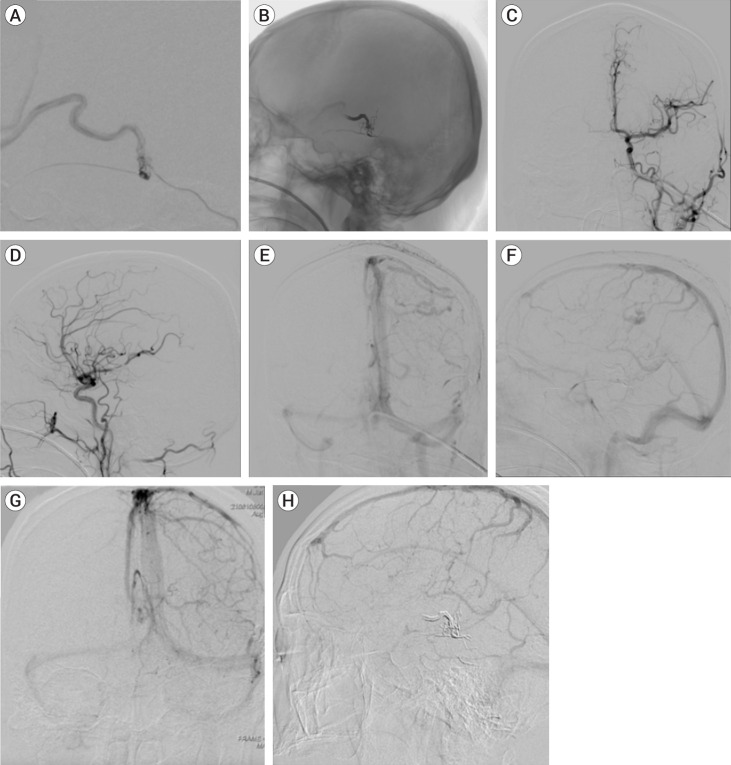
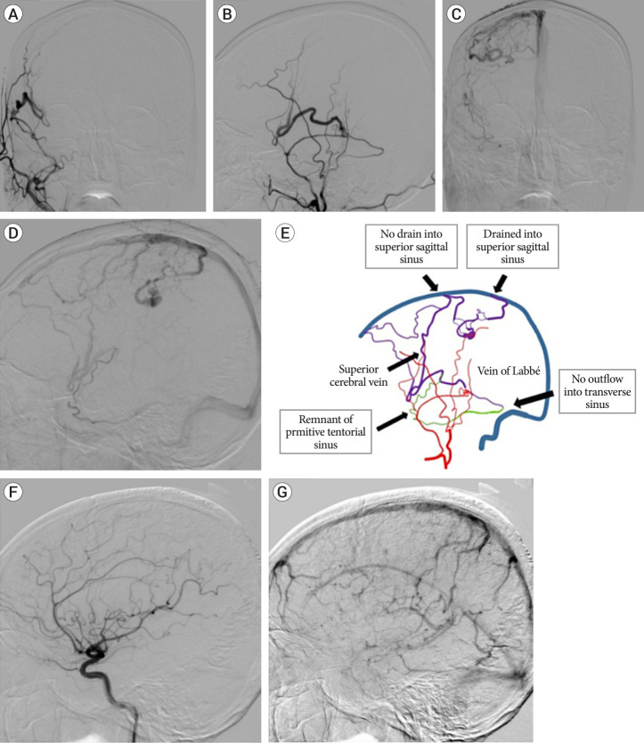
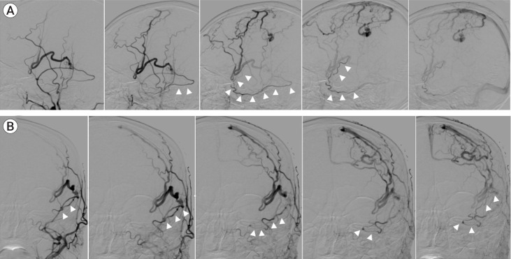
Convexity dural arteriovenous fistula (dAVF) is associated with high-grade dAVF and is usually presented with aggressive clinical presentation. Precise diagnosis and understanding the pathogenesis are important to achieving successful treatment without complications. We report a case of dAVF with Sylvian-Labbé collateral pattern, concerning embryological development that was thought to be involved in the vascular architecture and pathogenesis of dural AVF. Thus, a 60-year-old man was presented with sudden onset of seizure with no history of trauma. Magnetic Resonance Imaging (MRI) showed cortical hemorrhage in the left precentral gyrus. Digital subtraction angiography (DSA) showed the convexity dural arteriovenous fistula (dAVF) involving a vein that appeared to be the vein of Labbé, the drainer was anastomosed with superior middle cerebral vein (SMCV) and formed the varix. With the successful treatment with trans-arterial embolization (TAE), obliteration of dAVF was achieved with no neurological deficits. This case highlights convexity dAVF with the complex relationship between embryological development and the arcade of venous drainage route, wherein the anomaly might be acquired and caused by elevated venous pressure in a vein that appeared to be the vein of Labbé. Gaining knowledge of the embryological basis may aid in a deeper understanding of acquired pathologies.

 求助内容:
求助内容: 应助结果提醒方式:


