Antonio Carlos da S Senra Filho, Luiz Otávio Murta Junior, André Monteiro Paschoal
{"title":"Assessing biological self-organization patterns using statistical complexity characteristics: a tool for diffusion tensor imaging analysis.","authors":"Antonio Carlos da S Senra Filho, Luiz Otávio Murta Junior, André Monteiro Paschoal","doi":"10.1007/s10334-024-01185-4","DOIUrl":null,"url":null,"abstract":"<p><strong>Object: </strong>Diffusion-weighted imaging (DWI) and diffusion tensor imaging (DTI) are well-known and powerful imaging techniques for MRI. Although DTI evaluation has evolved continually in recent years, there are still struggles regarding quantitative measurements that can benefit brain areas that are consistently difficult to measure via diffusion-based methods, e.g., gray matter (GM). The present study proposes a new image processing technique based on diffusion distribution evaluation of López-Ruiz, Mancini and Calbet (LMC) complexity called diffusion complexity (DC).</p><p><strong>Materials and methods: </strong>The OASIS-3 and TractoInferno open-science databases for healthy individuals were used, and all the codes are provided as open-source materials.</p><p><strong>Results: </strong>The DC map showed relevant signal characterization in brain tissues and structures, achieving contrast-to-noise ratio (CNR) gains of approximately 39% and 93%, respectively, compared to those of the FA and ADC maps.</p><p><strong>Discussion: </strong>In the special case of GM tissue, the DC map obtains its maximum signal level, showing the possibility of studying cortical and subcortical structures challenging for classical DTI quantitative formalism. The ability to apply the DC technique, which requires the same imaging acquisition for DTI and its potential to provide complementary information to study the brain's GM structures, can be a rich source of information for further neuroscience research and clinical practice.</p>","PeriodicalId":18067,"journal":{"name":"Magnetic Resonance Materials in Physics, Biology and Medicine","volume":" ","pages":"653-663"},"PeriodicalIF":2.5000,"publicationDate":"2025-08-01","publicationTypes":"Journal Article","fieldsOfStudy":null,"isOpenAccess":false,"openAccessPdf":"","citationCount":"0","resultStr":null,"platform":"Semanticscholar","paperid":null,"PeriodicalName":"Magnetic Resonance Materials in Physics, Biology and Medicine","FirstCategoryId":"3","ListUrlMain":"https://doi.org/10.1007/s10334-024-01185-4","RegionNum":4,"RegionCategory":"医学","ArticlePicture":[],"TitleCN":null,"AbstractTextCN":null,"PMCID":null,"EPubDate":"2024/7/28 0:00:00","PubModel":"Epub","JCR":"Q3","JCRName":"RADIOLOGY, NUCLEAR MEDICINE & MEDICAL IMAGING","Score":null,"Total":0}
引用次数: 0
Abstract
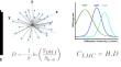
Object: Diffusion-weighted imaging (DWI) and diffusion tensor imaging (DTI) are well-known and powerful imaging techniques for MRI. Although DTI evaluation has evolved continually in recent years, there are still struggles regarding quantitative measurements that can benefit brain areas that are consistently difficult to measure via diffusion-based methods, e.g., gray matter (GM). The present study proposes a new image processing technique based on diffusion distribution evaluation of López-Ruiz, Mancini and Calbet (LMC) complexity called diffusion complexity (DC).
Materials and methods: The OASIS-3 and TractoInferno open-science databases for healthy individuals were used, and all the codes are provided as open-source materials.
Results: The DC map showed relevant signal characterization in brain tissues and structures, achieving contrast-to-noise ratio (CNR) gains of approximately 39% and 93%, respectively, compared to those of the FA and ADC maps.
Discussion: In the special case of GM tissue, the DC map obtains its maximum signal level, showing the possibility of studying cortical and subcortical structures challenging for classical DTI quantitative formalism. The ability to apply the DC technique, which requires the same imaging acquisition for DTI and its potential to provide complementary information to study the brain's GM structures, can be a rich source of information for further neuroscience research and clinical practice.
期刊介绍:
MAGMA is a multidisciplinary international journal devoted to the publication of articles on all aspects of magnetic resonance techniques and their applications in medicine and biology. MAGMA currently publishes research papers, reviews, letters to the editor, and commentaries, six times a year. The subject areas covered by MAGMA include:
advances in materials, hardware and software in magnetic resonance technology,
new developments and results in research and practical applications of magnetic resonance imaging and spectroscopy related to biology and medicine,
study of animal models and intact cells using magnetic resonance,
reports of clinical trials on humans and clinical validation of magnetic resonance protocols.

 求助内容:
求助内容: 应助结果提醒方式:


