{"title":"AQP4- and Kir4.1-Mediated Müller Cell Oedema Is Involved in Retinal Injury Induced By Hypobaric Hypoxia.","authors":"Cong Han, Yuting Li, Xingxing Zheng, Xiaoxia Zhang, Guonian Li, Liangtao Zhao, Zhaoqian Chen, Yi Yang, Wenfang Zhang","doi":"10.1007/s12035-024-04382-3","DOIUrl":null,"url":null,"abstract":"<p><p>Hypobaric hypoxia is the main cause of high-altitude retinopathy (HAR). Retinal oedema is the key pathological change in HAR. However, its pathological mechanism is not clear. In this study, a 5000-m hypobaric hypoxic environment was simulated. Haematoxylin and eosin (H&E) staining and electrophysiological (ERG) detection were used to observe the morphological and functional changes in the retina of mice under hypobaric hypoxia for 2-72 h. Toluidine blue staining and transmission electron microscopy were used to observe the morphology of Müller cells in the hypobaric hypoxia groups. The functional changes and oedema mechanism of Müller cells were detected by immunofluorescence and western blotting. The expression levels of glutamine synthetase (GS), glial fibrillary acidic protein (GFAP), aquaporin 4 (AQP4), and inwardly rectifying potassium channel subtype 4.1 (Kir4.1) in Müller cells were quantitatively analysed. This study revealed that retinal oedema gradually increased with prolonged exposure to a 5000-m hypobaric hypoxic environment. In addition, the ERG showed that the time delay and amplitude of the a-wave and b-wave decreased. The expression of GS decreased, and the expression of GFAP increased in Müller cells after exposure to hypobaric hypoxia for 4 h. At the same time, retinal AQP4 expression increased, and Kir4.1 expression decreased. The oedema and functional changes in Müller cells are consistent with the time point of retinal oedema. In conclusion, Müller cell oedema is involved in retinal oedema induced by hypobaric hypoxia. An increase in AQP4 and a decrease in Kir4.1 are the main causes of Müller cell oedema caused by hypobaric hypoxia.</p>","PeriodicalId":18762,"journal":{"name":"Molecular Neurobiology","volume":" ","pages":"2012-2022"},"PeriodicalIF":4.6000,"publicationDate":"2025-02-01","publicationTypes":"Journal Article","fieldsOfStudy":null,"isOpenAccess":false,"openAccessPdf":"","citationCount":"0","resultStr":null,"platform":"Semanticscholar","paperid":null,"PeriodicalName":"Molecular Neurobiology","FirstCategoryId":"3","ListUrlMain":"https://doi.org/10.1007/s12035-024-04382-3","RegionNum":2,"RegionCategory":"医学","ArticlePicture":[],"TitleCN":null,"AbstractTextCN":null,"PMCID":null,"EPubDate":"2024/7/26 0:00:00","PubModel":"Epub","JCR":"Q1","JCRName":"NEUROSCIENCES","Score":null,"Total":0}
引用次数: 0
Abstract
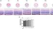
Hypobaric hypoxia is the main cause of high-altitude retinopathy (HAR). Retinal oedema is the key pathological change in HAR. However, its pathological mechanism is not clear. In this study, a 5000-m hypobaric hypoxic environment was simulated. Haematoxylin and eosin (H&E) staining and electrophysiological (ERG) detection were used to observe the morphological and functional changes in the retina of mice under hypobaric hypoxia for 2-72 h. Toluidine blue staining and transmission electron microscopy were used to observe the morphology of Müller cells in the hypobaric hypoxia groups. The functional changes and oedema mechanism of Müller cells were detected by immunofluorescence and western blotting. The expression levels of glutamine synthetase (GS), glial fibrillary acidic protein (GFAP), aquaporin 4 (AQP4), and inwardly rectifying potassium channel subtype 4.1 (Kir4.1) in Müller cells were quantitatively analysed. This study revealed that retinal oedema gradually increased with prolonged exposure to a 5000-m hypobaric hypoxic environment. In addition, the ERG showed that the time delay and amplitude of the a-wave and b-wave decreased. The expression of GS decreased, and the expression of GFAP increased in Müller cells after exposure to hypobaric hypoxia for 4 h. At the same time, retinal AQP4 expression increased, and Kir4.1 expression decreased. The oedema and functional changes in Müller cells are consistent with the time point of retinal oedema. In conclusion, Müller cell oedema is involved in retinal oedema induced by hypobaric hypoxia. An increase in AQP4 and a decrease in Kir4.1 are the main causes of Müller cell oedema caused by hypobaric hypoxia.
期刊介绍:
Molecular Neurobiology is an exciting journal for neuroscientists needing to stay in close touch with progress at the forefront of molecular brain research today. It is an especially important periodical for graduate students and "postdocs," specifically designed to synthesize and critically assess research trends for all neuroscientists hoping to stay active at the cutting edge of this dramatically developing area. This journal has proven to be crucial in departmental libraries, serving as essential reading for every committed neuroscientist who is striving to keep abreast of all rapid developments in a forefront field. Most recent significant advances in experimental and clinical neuroscience have been occurring at the molecular level. Until now, there has been no journal devoted to looking closely at this fragmented literature in a critical, coherent fashion. Each submission is thoroughly analyzed by scientists and clinicians internationally renowned for their special competence in the areas treated.
 求助内容:
求助内容: 应助结果提醒方式:


