Notochordal cell derived lesions: a 55-year casuistic analysis of 50 cases with radiologic-pathologic correlation in a tertiary referral hospital, and literature review.
Eva Manuela Pena-Burgos, Nerea Torena Lerchundi, Jorge Fuentes-Sánchez, Mar Tapia-Viñe, Nicomedes Fernández-Baíllo, Jose Juan Pozo-Kreilinger
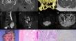
{"title":"Notochordal cell derived lesions: a 55-year casuistic analysis of 50 cases with radiologic-pathologic correlation in a tertiary referral hospital, and literature review.","authors":"Eva Manuela Pena-Burgos, Nerea Torena Lerchundi, Jorge Fuentes-Sánchez, Mar Tapia-Viñe, Nicomedes Fernández-Baíllo, Jose Juan Pozo-Kreilinger","doi":"10.1007/s00586-024-08419-y","DOIUrl":null,"url":null,"abstract":"<p><p>Distinct lesions are derived from notochordal cells (NCDL), ranging from benign to malignant ones. This study presents fifty NCDL cases diagnosed in a tertiary hospital of reference from the past 55 years: forty-two conventional chordomas, including one chondroid chordoma subtype, four benign notochordal cell tumors (BNCT), two conventional chordomas with BNCT foci, and two dedifferentiated chordomas. All patients were adults. Three BNCT were incidentally diagnosed, and one case presented local pain. Chordomas began with local pain and/or neurological symptoms. BNCT were well-defined intraosseous lesions, hypointense on T1-weighted images (WI) and hyperintense on T2-WI, without enhancement in the contrast. Conventional chordomas, including its chondroid subtype, were lobulated masses with cortical disruption and soft tissue extension, hypointense on T1-WI and hyperintense on T2-WI, with variable contrast enhancement. BNCT were histologically composed of solid sheets of vacuolated cells with clear cytoplasm and round and central nuclei. No atypia, lobular growth pattern, myxoid matrix, or bone infiltration were seen. Conventional chordomas were histologically composed of physaliphorous cells in a myxoid stroma with lobulated and infiltrating growth patterns. Observational follow-up using radiological controls was decided on for the BNCT cases. None of these cases presented local recurrence or metastasis. En-bloc resection and adjuvant radiotherapy were selected for sacral and vertebral chordoma cases. Sixteen patients died due to tumor-related factors; twenty-eight presented local recurrence, and four developed distant metastases. New therapeutic options are being studied for chordoma cases. Clinical, radiological, and histopathological data are necessary to properly diagnose and follow up of NCDL.</p>","PeriodicalId":12323,"journal":{"name":"European Spine Journal","volume":null,"pages":null},"PeriodicalIF":2.6000,"publicationDate":"2024-09-01","publicationTypes":"Journal Article","fieldsOfStudy":null,"isOpenAccess":false,"openAccessPdf":"","citationCount":"0","resultStr":null,"platform":"Semanticscholar","paperid":null,"PeriodicalName":"European Spine Journal","FirstCategoryId":"3","ListUrlMain":"https://doi.org/10.1007/s00586-024-08419-y","RegionNum":3,"RegionCategory":"医学","ArticlePicture":[],"TitleCN":null,"AbstractTextCN":null,"PMCID":null,"EPubDate":"2024/7/24 0:00:00","PubModel":"Epub","JCR":"Q2","JCRName":"CLINICAL NEUROLOGY","Score":null,"Total":0}
引用次数: 0
Abstract
Distinct lesions are derived from notochordal cells (NCDL), ranging from benign to malignant ones. This study presents fifty NCDL cases diagnosed in a tertiary hospital of reference from the past 55 years: forty-two conventional chordomas, including one chondroid chordoma subtype, four benign notochordal cell tumors (BNCT), two conventional chordomas with BNCT foci, and two dedifferentiated chordomas. All patients were adults. Three BNCT were incidentally diagnosed, and one case presented local pain. Chordomas began with local pain and/or neurological symptoms. BNCT were well-defined intraosseous lesions, hypointense on T1-weighted images (WI) and hyperintense on T2-WI, without enhancement in the contrast. Conventional chordomas, including its chondroid subtype, were lobulated masses with cortical disruption and soft tissue extension, hypointense on T1-WI and hyperintense on T2-WI, with variable contrast enhancement. BNCT were histologically composed of solid sheets of vacuolated cells with clear cytoplasm and round and central nuclei. No atypia, lobular growth pattern, myxoid matrix, or bone infiltration were seen. Conventional chordomas were histologically composed of physaliphorous cells in a myxoid stroma with lobulated and infiltrating growth patterns. Observational follow-up using radiological controls was decided on for the BNCT cases. None of these cases presented local recurrence or metastasis. En-bloc resection and adjuvant radiotherapy were selected for sacral and vertebral chordoma cases. Sixteen patients died due to tumor-related factors; twenty-eight presented local recurrence, and four developed distant metastases. New therapeutic options are being studied for chordoma cases. Clinical, radiological, and histopathological data are necessary to properly diagnose and follow up of NCDL.
期刊介绍:
"European Spine Journal" is a publication founded in response to the increasing trend toward specialization in spinal surgery and spinal pathology in general. The Journal is devoted to all spine related disciplines, including functional and surgical anatomy of the spine, biomechanics and pathophysiology, diagnostic procedures, and neurology, surgery and outcomes. The aim of "European Spine Journal" is to support the further development of highly innovative spine treatments including but not restricted to surgery and to provide an integrated and balanced view of diagnostic, research and treatment procedures as well as outcomes that will enhance effective collaboration among specialists worldwide. The “European Spine Journal” also participates in education by means of videos, interactive meetings and the endorsement of educative efforts.
Official publication of EUROSPINE, The Spine Society of Europe

 求助内容:
求助内容: 应助结果提醒方式:


