Development and first implementation of a novel multi-modality cardiac motion and dosimetry phantom for radiotherapy applications

IF 3.2 2区 医学 Q1 RADIOLOGY, NUCLEAR MEDICINE & MEDICAL IMAGING
Medical physics Pub Date : 2024-07-23 DOI:10.1002/mp.17315
Kenneth W. Gregg, Chase Ruff, Grant Koenig, Kalin I. Penev, Andrew Shepard, Grace Kreissler, Margo Amatuzio, Cameron Owens, Prashant Nagpal, Carri K. Glide-Hurst
{"title":"Development and first implementation of a novel multi-modality cardiac motion and dosimetry phantom for radiotherapy applications","authors":"Kenneth W. Gregg,&nbsp;Chase Ruff,&nbsp;Grant Koenig,&nbsp;Kalin I. Penev,&nbsp;Andrew Shepard,&nbsp;Grace Kreissler,&nbsp;Margo Amatuzio,&nbsp;Cameron Owens,&nbsp;Prashant Nagpal,&nbsp;Carri K. Glide-Hurst","doi":"10.1002/mp.17315","DOIUrl":null,"url":null,"abstract":"<div>\n \n \n <section>\n \n <h3> Background</h3>\n \n <p>Cardiac applications in radiation therapy are rapidly expanding including magnetic resonance guided radiation therapy (MRgRT) for real-time gating for targeting and avoidance near the heart or treating ventricular tachycardia (VT).</p>\n </section>\n \n <section>\n \n <h3> Purpose</h3>\n \n <p>This work describes the development and implementation of a novel multi-modality and magnetic resonance (MR)-compatible cardiac phantom.</p>\n </section>\n \n <section>\n \n <h3> Methods</h3>\n \n <p>The patient-informed 3D model was derived from manual contouring of a contrast-enhanced Coronary Computed Tomography Angiography scan, exported as a Stereolithography model, then post-processed to simulate female heart with an average volume. The model was 3D-printed using Elastic50A to provide MR contrast to water background. Two rigid acrylic modules containing cardiac structures were designed and assembled, retrofitting to an MR-safe programmable motor to supply cardiac and respiratory motion in superior-inferior directions. One module contained a cavity for an ion chamber (IC), and the other was equipped with multiple interchangeable cavities for plastic scintillation detectors (PSDs). Images were acquired on a 0.35 T MR-linac for validation of phantom geometry, motion, and simulated online treatment planning and delivery. Three motion profiles were prescribed: patient-derived cardiac (sine waveform, 4.3 mm peak-to-peak, 60 beats/min), respiratory (cos<sup>4</sup> waveform, 30 mm peak-to-peak, 12 breaths/min), and a superposition of cardiac (sine waveform, 4 mm peak-to-peak, 70 beats/min) and respiratory (cos<sup>4</sup> waveform, 24 mm peak-to-peak, 12 breaths/min). The amplitude of the motion profiles was evaluated from sagittal cine images at eight frames/s with a resolution of 2.4 mm × 2.4 mm. Gated dosimetry experiments were performed using the two module configurations for calculating dose relative to stationary. A CT-based VT treatment plan was delivered twice under cone-beam CT guidance and cumulative stationary doses to multi-point PSDs were evaluated.</p>\n </section>\n \n <section>\n \n <h3> Results</h3>\n \n <p>No artifacts were observed on any images acquired during phantom operation. Phantom excursions measured 49.3 ± 25.8%/66.9 ± 14.0%, 97.0 ± 2.2%/96.4 ± 1.7%, and 90.4 ± 4.8%/89.3 ± 3.5% of prescription for cardiac, respiratory, and cardio-respiratory motion profiles for the 2-chamber (PSD) and 12-substructure (IC) phantom modules respectively. In the gated experiments, the cumulative dose was &lt;2% from expected using the IC module. Real-time dose measured for the PSDs at 10 Hz acquisition rate demonstrated the ability to detect the dosimetric consequences of cardiac, respiratory, and cardio-respiratory motion when sampling of different locations during a single delivery, and the stability of our phantom dosimetric results over repeated cycles for the high dose and high gradient regions. For the VT delivery, high dose PSD was &lt;1% from expected (5–6 cGy deviation of 5.9 Gy/fraction) and high gradient/low dose regions had deviations &lt;3.6% (6.3 cGy less than expected 1.73 Gy/fraction).</p>\n </section>\n \n <section>\n \n <h3> Conclusions</h3>\n \n <p>A novel multi-modality modular heart phantom was designed, constructed, and used for gated radiotherapy experiments on a 0.35 T MR-linac. Our phantom was capable of mimicking cardiac, cardio-respiratory, and respiratory motion while performing dosimetric evaluations of gated procedures using IC and PSD configurations. Time-resolved PSDs with small sensitive volumes appear promising for low-amplitude/high-frequency motion and multi-point data acquisition for advanced dosimetric capabilities. Illustrating VT planning and delivery further expands our phantom to address the unmet needs of cardiac applications in radiotherapy.</p>\n </section>\n </div>","PeriodicalId":18384,"journal":{"name":"Medical physics","volume":"51 10","pages":"7479-7491"},"PeriodicalIF":3.2000,"publicationDate":"2024-07-23","publicationTypes":"Journal Article","fieldsOfStudy":null,"isOpenAccess":false,"openAccessPdf":"https://onlinelibrary.wiley.com/doi/epdf/10.1002/mp.17315","citationCount":"0","resultStr":null,"platform":"Semanticscholar","paperid":null,"PeriodicalName":"Medical physics","FirstCategoryId":"3","ListUrlMain":"https://onlinelibrary.wiley.com/doi/10.1002/mp.17315","RegionNum":2,"RegionCategory":"医学","ArticlePicture":[],"TitleCN":null,"AbstractTextCN":null,"PMCID":null,"EPubDate":"","PubModel":"","JCR":"Q1","JCRName":"RADIOLOGY, NUCLEAR MEDICINE & MEDICAL IMAGING","Score":null,"Total":0}
引用次数: 0

Abstract

Background

Cardiac applications in radiation therapy are rapidly expanding including magnetic resonance guided radiation therapy (MRgRT) for real-time gating for targeting and avoidance near the heart or treating ventricular tachycardia (VT).

Purpose

This work describes the development and implementation of a novel multi-modality and magnetic resonance (MR)-compatible cardiac phantom.

Methods

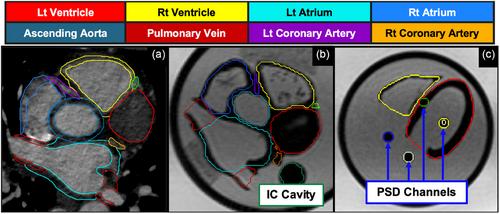
The patient-informed 3D model was derived from manual contouring of a contrast-enhanced Coronary Computed Tomography Angiography scan, exported as a Stereolithography model, then post-processed to simulate female heart with an average volume. The model was 3D-printed using Elastic50A to provide MR contrast to water background. Two rigid acrylic modules containing cardiac structures were designed and assembled, retrofitting to an MR-safe programmable motor to supply cardiac and respiratory motion in superior-inferior directions. One module contained a cavity for an ion chamber (IC), and the other was equipped with multiple interchangeable cavities for plastic scintillation detectors (PSDs). Images were acquired on a 0.35 T MR-linac for validation of phantom geometry, motion, and simulated online treatment planning and delivery. Three motion profiles were prescribed: patient-derived cardiac (sine waveform, 4.3 mm peak-to-peak, 60 beats/min), respiratory (cos4 waveform, 30 mm peak-to-peak, 12 breaths/min), and a superposition of cardiac (sine waveform, 4 mm peak-to-peak, 70 beats/min) and respiratory (cos4 waveform, 24 mm peak-to-peak, 12 breaths/min). The amplitude of the motion profiles was evaluated from sagittal cine images at eight frames/s with a resolution of 2.4 mm × 2.4 mm. Gated dosimetry experiments were performed using the two module configurations for calculating dose relative to stationary. A CT-based VT treatment plan was delivered twice under cone-beam CT guidance and cumulative stationary doses to multi-point PSDs were evaluated.

Results

No artifacts were observed on any images acquired during phantom operation. Phantom excursions measured 49.3 ± 25.8%/66.9 ± 14.0%, 97.0 ± 2.2%/96.4 ± 1.7%, and 90.4 ± 4.8%/89.3 ± 3.5% of prescription for cardiac, respiratory, and cardio-respiratory motion profiles for the 2-chamber (PSD) and 12-substructure (IC) phantom modules respectively. In the gated experiments, the cumulative dose was <2% from expected using the IC module. Real-time dose measured for the PSDs at 10 Hz acquisition rate demonstrated the ability to detect the dosimetric consequences of cardiac, respiratory, and cardio-respiratory motion when sampling of different locations during a single delivery, and the stability of our phantom dosimetric results over repeated cycles for the high dose and high gradient regions. For the VT delivery, high dose PSD was <1% from expected (5–6 cGy deviation of 5.9 Gy/fraction) and high gradient/low dose regions had deviations <3.6% (6.3 cGy less than expected 1.73 Gy/fraction).

Conclusions

A novel multi-modality modular heart phantom was designed, constructed, and used for gated radiotherapy experiments on a 0.35 T MR-linac. Our phantom was capable of mimicking cardiac, cardio-respiratory, and respiratory motion while performing dosimetric evaluations of gated procedures using IC and PSD configurations. Time-resolved PSDs with small sensitive volumes appear promising for low-amplitude/high-frequency motion and multi-point data acquisition for advanced dosimetric capabilities. Illustrating VT planning and delivery further expands our phantom to address the unmet needs of cardiac applications in radiotherapy.
Abstract Image

开发并首次实施用于放射治疗的新型多模态心脏运动和剂量测定模型。
背景:心脏放射治疗的应用范围正在迅速扩大,包括磁共振引导放射治疗(MRgRT),用于实时选取目标、避开心脏附近或治疗室性心动过速(VT)。目的:这项工作描述了新型多模态和磁共振(MR)兼容心脏模型的开发和实施:患者信息三维模型来自对比增强冠状动脉计算机断层扫描血管造影扫描的人工轮廓,导出为立体光刻模型,然后进行后处理以模拟具有平均体积的女性心脏。该模型使用 Elastic50A 进行 3D 打印,以提供磁共振成像与水背景的对比度。设计并组装了两个包含心脏结构的硬质丙烯酸模块,并加装了磁共振安全可编程电机,以提供上下方向的心脏和呼吸运动。其中一个模块包含一个离子室(IC)空腔,另一个模块则配备了多个可互换的塑料闪烁探测器(PSD)空腔。在 0.35 T MR-linac 上采集图像,以验证模型的几何形状、运动以及模拟在线治疗计划和实施。我们设定了三种运动曲线:患者来源的心脏运动曲线(正弦波形,4.3 毫米峰峰值,60 次/分钟)、呼吸运动曲线(cos4 波形,30 毫米峰峰值,12 次/分钟)以及心脏运动曲线(正弦波形,4 毫米峰峰值,70 次/分钟)和呼吸运动曲线(cos4 波形,24 毫米峰峰值,12 次/分钟)的叠加。运动轮廓的振幅是通过分辨率为 2.4 毫米 × 2.4 毫米的矢状弧线图像以每秒八帧的速度进行评估的。使用两种模块配置进行了门控剂量测定实验,以计算相对于静态的剂量。在锥形束 CT 引导下,进行了两次基于 CT 的 VT 治疗计划,并对多点 PSD 的累积固定剂量进行了评估:结果:在模型运行期间采集的任何图像上都没有发现伪影。两腔(PSD)和 12 子结构(IC)模型模块的心脏、呼吸和心肺运动曲线的偏移量分别为 49.3 ± 25.8%/66.9 ± 14.0%、97.0 ± 2.2%/96.4 ± 1.7%,以及 90.4 ± 4.8%/89.3 ± 3.5%。在门控实验中,累积剂量为结论:我们设计、制作了一种新型多模态模块化心脏模型,并将其用于 0.35 T 磁共振成像仪上的选通放疗实验。我们的模型能够模拟心脏、心肺和呼吸运动,同时使用 IC 和 PSD 配置对门控程序进行剂量学评估。具有小敏感体积的时间分辨 PSD 对于低振幅/高频运动和先进剂量测定能力的多点数据采集很有前景。说明 VT 计划和传输进一步扩展了我们的模型,以满足放疗中未得到满足的心脏应用需求。
本文章由计算机程序翻译,如有差异,请以英文原文为准。
求助全文
约1分钟内获得全文 求助全文
来源期刊
Medical physics
Medical physics 医学-核医学
CiteScore
6.80
自引率
15.80%
发文量
660
审稿时长
1.7 months
期刊介绍: Medical Physics publishes original, high impact physics, imaging science, and engineering research that advances patient diagnosis and therapy through contributions in 1) Basic science developments with high potential for clinical translation 2) Clinical applications of cutting edge engineering and physics innovations 3) Broadly applicable and innovative clinical physics developments Medical Physics is a journal of global scope and reach. By publishing in Medical Physics your research will reach an international, multidisciplinary audience including practicing medical physicists as well as physics- and engineering based translational scientists. We work closely with authors of promising articles to improve their quality.
×
引用
GB/T 7714-2015
复制
MLA
复制
APA
复制
导出至
BibTeX EndNote RefMan NoteFirst NoteExpress
×
提示
您的信息不完整,为了账户安全,请先补充。
现在去补充
×
提示
您因"违规操作"
具体请查看互助需知
我知道了
×
提示
确定
请完成安全验证×
copy
已复制链接
快去分享给好友吧!
我知道了
右上角分享
点击右上角分享
0
联系我们:info@booksci.cn Book学术提供免费学术资源搜索服务,方便国内外学者检索中英文文献。致力于提供最便捷和优质的服务体验。 Copyright © 2023 布克学术 All rights reserved.
京ICP备2023020795号-1
ghs 京公网安备 11010802042870号
Book学术文献互助
Book学术文献互助群
群 号:481959085
Book学术官方微信