{"title":"Tendinous Signal Alterations on MRI in the Asymptomatic Elbow: A Retrospective Cross-Sectional Study.","authors":"Bjorn Valgaeren, Elyn Van Snick, Bart Claikens","doi":"10.5334/jbsr.3651","DOIUrl":null,"url":null,"abstract":"<p><strong>Objectives: </strong>It is clinically relevant to prevent overtreatment of tendinopathy diagnosed solely on imaging. Therefore, the prevalence of presumable asymptomatic signal changes in the common flexor origin, biceps insertion, brachialis insertion, and triceps insertion were assessed.</p><p><strong>Materials and methods: </strong>Two hundred and five magnetic resonance imaging (MRI) exams of the elbow with coronal and axial fat-saturated fluid-sensitive sequences between January 1, 2018 and July 31, 2022 were retrospectively identified in our center.Two radiology residents reviewed the exams independently. The elbow tendons were given a score from 0 to 4. Score 0: no signal abnormality; score 1: increased T2-weighted signal around the tendon; score 2: increased T2-weighted signal compared to muscle within the tendon; score 3: partial tear; and score 4: complete tear.</p><p><strong>Results: </strong>The common flexor tendon showed signal alterations in 8% of patients; nine patients had an increased signal around the tendon, and eight patients had an increased signal within the tendon. Three patients (1.5%) had an altered signal intensity in the biceps tendon. All triceps tendons showed a linear hyperintense signal, suggesting that it is physiological. There were no partial or complete tears. No signal abnormalities were noted in the brachialis tendon among all patients.</p><p><strong>Conclusion: </strong>The prevalence of presumable asymptomatic signal alterations seen in the common flexor origin on MRI is not negligible; therefore, clinical correlation is advised to prevent overtreatment of tendinopathy in these cases. No partial or complete tears were seen.</p>","PeriodicalId":55987,"journal":{"name":"Journal of the Belgian Society of Radiology","volume":"108 1","pages":"69"},"PeriodicalIF":1.3000,"publicationDate":"2024-07-13","publicationTypes":"Journal Article","fieldsOfStudy":null,"isOpenAccess":false,"openAccessPdf":"https://www.ncbi.nlm.nih.gov/pmc/articles/PMC11259101/pdf/","citationCount":"0","resultStr":null,"platform":"Semanticscholar","paperid":null,"PeriodicalName":"Journal of the Belgian Society of Radiology","FirstCategoryId":"3","ListUrlMain":"https://doi.org/10.5334/jbsr.3651","RegionNum":4,"RegionCategory":"医学","ArticlePicture":[],"TitleCN":null,"AbstractTextCN":null,"PMCID":null,"EPubDate":"2024/1/1 0:00:00","PubModel":"eCollection","JCR":"Q4","JCRName":"RADIOLOGY, NUCLEAR MEDICINE & MEDICAL IMAGING","Score":null,"Total":0}
引用次数: 0
Abstract
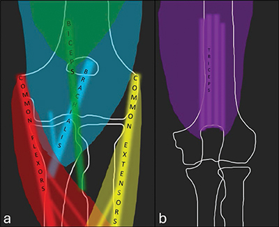
Objectives: It is clinically relevant to prevent overtreatment of tendinopathy diagnosed solely on imaging. Therefore, the prevalence of presumable asymptomatic signal changes in the common flexor origin, biceps insertion, brachialis insertion, and triceps insertion were assessed.
Materials and methods: Two hundred and five magnetic resonance imaging (MRI) exams of the elbow with coronal and axial fat-saturated fluid-sensitive sequences between January 1, 2018 and July 31, 2022 were retrospectively identified in our center.Two radiology residents reviewed the exams independently. The elbow tendons were given a score from 0 to 4. Score 0: no signal abnormality; score 1: increased T2-weighted signal around the tendon; score 2: increased T2-weighted signal compared to muscle within the tendon; score 3: partial tear; and score 4: complete tear.
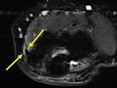
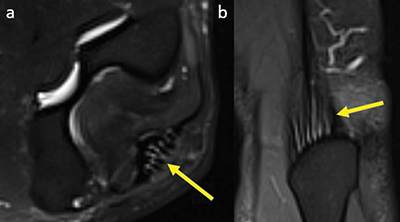
Results: The common flexor tendon showed signal alterations in 8% of patients; nine patients had an increased signal around the tendon, and eight patients had an increased signal within the tendon. Three patients (1.5%) had an altered signal intensity in the biceps tendon. All triceps tendons showed a linear hyperintense signal, suggesting that it is physiological. There were no partial or complete tears. No signal abnormalities were noted in the brachialis tendon among all patients.
Conclusion: The prevalence of presumable asymptomatic signal alterations seen in the common flexor origin on MRI is not negligible; therefore, clinical correlation is advised to prevent overtreatment of tendinopathy in these cases. No partial or complete tears were seen.
期刊介绍:
The purpose of the Journal of the Belgian Society of Radiology is the publication of articles dealing with diagnostic and interventional radiology, related imaging techniques, allied sciences, and continuing education.
 求助内容:
求助内容: 应助结果提醒方式:


