Shunsuke Kasai, Akio Shiomi, Hideyuki Shimizu, Monami Aoba, Yusuke Kinugasa, Takuya Miura, Kay Uehara, Jun Watanabe, Kazushige Kawai, Yoichi Ajioka
{"title":"Risk factors and development of machine learning diagnostic models for lateral lymph node metastasis in rectal cancer: multicentre study.","authors":"Shunsuke Kasai, Akio Shiomi, Hideyuki Shimizu, Monami Aoba, Yusuke Kinugasa, Takuya Miura, Kay Uehara, Jun Watanabe, Kazushige Kawai, Yoichi Ajioka","doi":"10.1093/bjsopen/zrae073","DOIUrl":null,"url":null,"abstract":"<p><strong>Background: </strong>The diagnostic criteria for lateral lymph node metastasis in rectal cancer have not been established. This research aimed to investigate the risk factors for lateral lymph node metastasis and develop machine learning models combining these risk factors to improve the diagnostic performance of standard imaging.</p><p><strong>Method: </strong>This multicentre prospective study included patients who underwent lateral lymph node dissection without preoperative treatment for rectal cancer between 2017 and 2019 in 15 Japanese institutions. First, preoperative clinicopathological factors and magnetic resonance imaging findings were evaluated using multivariable analyses for their correlation with lateral lymph node metastasis. Next, machine learning diagnostic models for lateral lymph node metastasis were developed combining these risk factors. The models were tested in a training set and in an internal validation cohort and their diagnostic performance was tested using receiver operating characteristic curve analyses.</p><p><strong>Results: </strong>Of 212 rectal cancers, 122 patients were selected, including 232 lateral pelvic sides, 30 sides of which had pathological lateral lymph node metastasis. Multivariable analysis revealed that poorly differentiated/mucinous adenocarcinoma, extramural vascular invasion, tumour deposit and a short-axis diameter of lateral lymph node ≥ 6.0 mm were independent risk factors for lateral lymph node metastasis. Patients were randomly divided into a training cohort (139 sides) and a test cohort (93 sides) and machine learning models were computed on the basis of a combination of significant features (including: histological type, extramural vascular invasion, tumour deposit, short- and long-axis diameter of lateral lymph node, body mass index, serum carcinoembryonic antigen level, cT, cN, cM, irregular border and mixed signal intensity). The top three models with the highest sensitivity in the training cohort were as follows: support vector machine (sensitivity, 1.000; specificity, 0.773), light gradient boosting machine (sensitivity, 0.950; specificity, 0.918) and ensemble learning (sensitivity, 0.950; specificity, 0.917). The diagnostic performances of these models in the test cohort were as follows: support vector machine (sensitivity, 0.750; specificity, 0.667), light gradient boosting machine (sensitivity, 0.500; specificity, 0.852) and ensemble learning (sensitivity, 0.667; specificity, 0.864).</p><p><strong>Conclusion: </strong>Machine learning models combining multiple risk factors can contribute to improving diagnostic performance of lateral lymph node metastasis.</p>","PeriodicalId":9028,"journal":{"name":"BJS Open","volume":"8 4","pages":""},"PeriodicalIF":4.5000,"publicationDate":"2024-07-02","publicationTypes":"Journal Article","fieldsOfStudy":null,"isOpenAccess":false,"openAccessPdf":"https://www.ncbi.nlm.nih.gov/pmc/articles/PMC11252850/pdf/","citationCount":"0","resultStr":null,"platform":"Semanticscholar","paperid":null,"PeriodicalName":"BJS Open","FirstCategoryId":"3","ListUrlMain":"https://doi.org/10.1093/bjsopen/zrae073","RegionNum":3,"RegionCategory":"医学","ArticlePicture":[],"TitleCN":null,"AbstractTextCN":null,"PMCID":null,"EPubDate":"","PubModel":"","JCR":"Q1","JCRName":"SURGERY","Score":null,"Total":0}
引用次数: 0
Abstract
Background: The diagnostic criteria for lateral lymph node metastasis in rectal cancer have not been established. This research aimed to investigate the risk factors for lateral lymph node metastasis and develop machine learning models combining these risk factors to improve the diagnostic performance of standard imaging.
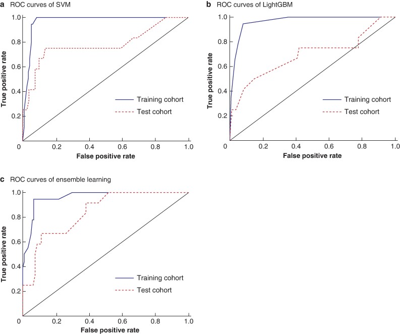
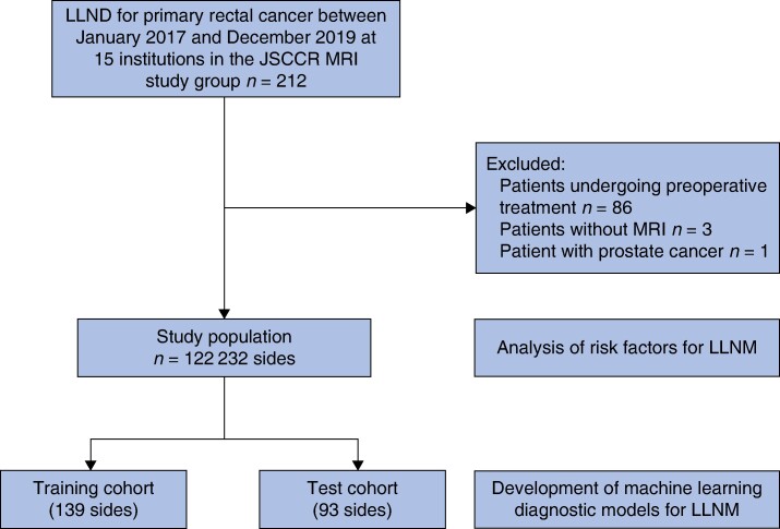
Method: This multicentre prospective study included patients who underwent lateral lymph node dissection without preoperative treatment for rectal cancer between 2017 and 2019 in 15 Japanese institutions. First, preoperative clinicopathological factors and magnetic resonance imaging findings were evaluated using multivariable analyses for their correlation with lateral lymph node metastasis. Next, machine learning diagnostic models for lateral lymph node metastasis were developed combining these risk factors. The models were tested in a training set and in an internal validation cohort and their diagnostic performance was tested using receiver operating characteristic curve analyses.
Results: Of 212 rectal cancers, 122 patients were selected, including 232 lateral pelvic sides, 30 sides of which had pathological lateral lymph node metastasis. Multivariable analysis revealed that poorly differentiated/mucinous adenocarcinoma, extramural vascular invasion, tumour deposit and a short-axis diameter of lateral lymph node ≥ 6.0 mm were independent risk factors for lateral lymph node metastasis. Patients were randomly divided into a training cohort (139 sides) and a test cohort (93 sides) and machine learning models were computed on the basis of a combination of significant features (including: histological type, extramural vascular invasion, tumour deposit, short- and long-axis diameter of lateral lymph node, body mass index, serum carcinoembryonic antigen level, cT, cN, cM, irregular border and mixed signal intensity). The top three models with the highest sensitivity in the training cohort were as follows: support vector machine (sensitivity, 1.000; specificity, 0.773), light gradient boosting machine (sensitivity, 0.950; specificity, 0.918) and ensemble learning (sensitivity, 0.950; specificity, 0.917). The diagnostic performances of these models in the test cohort were as follows: support vector machine (sensitivity, 0.750; specificity, 0.667), light gradient boosting machine (sensitivity, 0.500; specificity, 0.852) and ensemble learning (sensitivity, 0.667; specificity, 0.864).
Conclusion: Machine learning models combining multiple risk factors can contribute to improving diagnostic performance of lateral lymph node metastasis.

 求助内容:
求助内容: 应助结果提醒方式:


