The morphological discrepancy of neuromuscular junctions between bilateral paraspinal muscles in patients with adolescent idiopathic scoliosis: A quantitative immunofluorescence assay

IF 3.4 3区 医学 Q1 ORTHOPEDICS
JOR Spine Pub Date : 2024-07-15 DOI:10.1002/jsp2.1358
Tianyuan Zhang, Wenyuan Sui, Bin Li, Xiexiang Shao, Yaolong Deng, Zifang Zhang, Jingfan Yang, Zifang Huang, Wenjun Yang, Junlin Yang
{"title":"The morphological discrepancy of neuromuscular junctions between bilateral paraspinal muscles in patients with adolescent idiopathic scoliosis: A quantitative immunofluorescence assay","authors":"Tianyuan Zhang,&nbsp;Wenyuan Sui,&nbsp;Bin Li,&nbsp;Xiexiang Shao,&nbsp;Yaolong Deng,&nbsp;Zifang Zhang,&nbsp;Jingfan Yang,&nbsp;Zifang Huang,&nbsp;Wenjun Yang,&nbsp;Junlin Yang","doi":"10.1002/jsp2.1358","DOIUrl":null,"url":null,"abstract":"<div>\n \n \n <section>\n \n <h3> Introduction</h3>\n \n <p>Prior studies suggested that neuromuscular factors might be involved in the pathogenesis of adolescent idiopathic scoliosis (AIS). The neuromuscular junction (NMJ) is the important pivot where the nervous system interacts with muscle fibers, but it has not been well characterized in the paraspinal muscles of AIS. This study aims to perform the quantitative morphological analysis of NMJs from paraspinal muscles of AIS.</p>\n </section>\n \n <section>\n \n <h3> Methods</h3>\n \n <p>AIS patients who received surgery in our center were prospectively enrolled. Meanwhile, age-matched congenital scoliosis (CS) and non-scoliosis patients were also included as controls. Fresh samples of paraspinal muscles were harvested intraoperatively. NMJs were immunolabeled using different antibodies to reveal pre-synaptic neuronal architecture and post-synaptic motor endplates. A confocal microscope was used to acquire z-stack projections of NMJs images. Then, NMJs images were analyzed on maximum intensity projections using ImageJ software. The morphology of NMJs was quantitatively measured by a standardized ‘NMJ-morph’ workflow. A total of 21 variables were measured and compared between different groups.</p>\n </section>\n \n <section>\n \n <h3> Results</h3>\n \n <p>A total of 15 AIS patients, 10 CS patients and 5 normal controls were enrolled initially. For AIS group, NMJs in the convex side of paraspinal muscles demonstrated obviously decreased overlap when compared with the concave side (34.27% ± 8.09% vs. 48.11% ± 10.31%, <i>p</i> = 0.0036). However, no variables showed statistical difference between both sides of paraspinal muscles in CS patients. In contrast with non-scoliosis controls, both sides of paraspinal muscles in AIS patients demonstrated significantly smaller muscle bundle diameters.</p>\n </section>\n \n <section>\n \n <h3> Conclusions</h3>\n \n <p>This study first elucidated the morphological features of NMJs from paraspinal muscles of AIS patients. The NMJs in the convex side showed smaller overlap for AIS patients, but no difference was found in CS. This proved further evidence that neuromuscular factors might contribute to the mechanisms of AIS and could be considered as a novel potential therapeutic target for the treatment of progressive AIS.</p>\n </section>\n </div>","PeriodicalId":14876,"journal":{"name":"JOR Spine","volume":"7 3","pages":""},"PeriodicalIF":3.4000,"publicationDate":"2024-07-15","publicationTypes":"Journal Article","fieldsOfStudy":null,"isOpenAccess":false,"openAccessPdf":"https://www.ncbi.nlm.nih.gov/pmc/articles/PMC11247395/pdf/","citationCount":"0","resultStr":null,"platform":"Semanticscholar","paperid":null,"PeriodicalName":"JOR Spine","FirstCategoryId":"3","ListUrlMain":"https://onlinelibrary.wiley.com/doi/10.1002/jsp2.1358","RegionNum":3,"RegionCategory":"医学","ArticlePicture":[],"TitleCN":null,"AbstractTextCN":null,"PMCID":null,"EPubDate":"","PubModel":"","JCR":"Q1","JCRName":"ORTHOPEDICS","Score":null,"Total":0}
引用次数: 0

Abstract

Introduction

Prior studies suggested that neuromuscular factors might be involved in the pathogenesis of adolescent idiopathic scoliosis (AIS). The neuromuscular junction (NMJ) is the important pivot where the nervous system interacts with muscle fibers, but it has not been well characterized in the paraspinal muscles of AIS. This study aims to perform the quantitative morphological analysis of NMJs from paraspinal muscles of AIS.

Methods

AIS patients who received surgery in our center were prospectively enrolled. Meanwhile, age-matched congenital scoliosis (CS) and non-scoliosis patients were also included as controls. Fresh samples of paraspinal muscles were harvested intraoperatively. NMJs were immunolabeled using different antibodies to reveal pre-synaptic neuronal architecture and post-synaptic motor endplates. A confocal microscope was used to acquire z-stack projections of NMJs images. Then, NMJs images were analyzed on maximum intensity projections using ImageJ software. The morphology of NMJs was quantitatively measured by a standardized ‘NMJ-morph’ workflow. A total of 21 variables were measured and compared between different groups.

Results

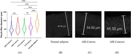
A total of 15 AIS patients, 10 CS patients and 5 normal controls were enrolled initially. For AIS group, NMJs in the convex side of paraspinal muscles demonstrated obviously decreased overlap when compared with the concave side (34.27% ± 8.09% vs. 48.11% ± 10.31%, p = 0.0036). However, no variables showed statistical difference between both sides of paraspinal muscles in CS patients. In contrast with non-scoliosis controls, both sides of paraspinal muscles in AIS patients demonstrated significantly smaller muscle bundle diameters.

Conclusions

This study first elucidated the morphological features of NMJs from paraspinal muscles of AIS patients. The NMJs in the convex side showed smaller overlap for AIS patients, but no difference was found in CS. This proved further evidence that neuromuscular factors might contribute to the mechanisms of AIS and could be considered as a novel potential therapeutic target for the treatment of progressive AIS.
Abstract Image

青少年特发性脊柱侧凸患者双侧脊柱旁肌肉神经肌肉接头的形态差异:免疫荧光定量分析。
导言:先前的研究表明,神经肌肉因素可能与青少年特发性脊柱侧弯症(AIS)的发病机制有关。神经肌肉接头(NMJ)是神经系统与肌肉纤维相互作用的重要枢纽,但在 AIS 的脊柱旁肌肉中尚未得到很好的表征。本研究旨在对AIS脊柱旁肌肉的NMJ进行定量形态学分析:方法:前瞻性地纳入在本中心接受手术的 AIS 患者。方法:前瞻性纳入在本中心接受手术的 AIS 患者,同时纳入年龄匹配的先天性脊柱侧凸(CS)和非脊柱侧凸患者作为对照。术中采集脊柱旁肌肉的新鲜样本。使用不同的抗体对NMJ进行免疫标记,以显示突触前神经元结构和突触后运动终板。使用共聚焦显微镜获取 NMJs 图像的 Z 叠投影。然后,使用 ImageJ 软件对最大强度投影的 NMJs 图像进行分析。通过标准化的 "NMJ-morph "工作流程对 NMJ 的形态进行定量测量。共测量了 21 个变量,并在不同组间进行了比较:结果:最初共招募了 15 名 AIS 患者、10 名 CS 患者和 5 名正常对照组。在 AIS 组中,脊柱旁肌肉凸面的 NMJ 重叠率明显低于凹面(34.27% ± 8.09% vs. 48.11% ± 10.31%,P = 0.0036)。然而,CS 患者脊柱旁肌肉两侧的变量均无统计学差异。与非脊柱侧弯对照组相比,AIS 患者两侧脊柱旁肌肉的肌束直径明显较小:本研究首次阐明了 AIS 患者脊柱旁肌肉 NMJ 的形态特征。AIS 患者凸侧的 NMJ 重叠较小,而 CS 患者则无差异。这进一步证明了神经肌肉因素可能是 AIS 的发病机制之一,并可被视为治疗进行性 AIS 的新的潜在治疗靶点。
本文章由计算机程序翻译,如有差异,请以英文原文为准。
求助全文
约1分钟内获得全文 求助全文
来源期刊
JOR Spine
JOR Spine ORTHOPEDICS-
CiteScore
6.40
自引率
18.90%
发文量
42
审稿时长
10 weeks
×
引用
GB/T 7714-2015
复制
MLA
复制
APA
复制
导出至
BibTeX EndNote RefMan NoteFirst NoteExpress
×
提示
您的信息不完整,为了账户安全,请先补充。
现在去补充
×
提示
您因"违规操作"
具体请查看互助需知
我知道了
×
提示
确定
请完成安全验证×
copy
已复制链接
快去分享给好友吧!
我知道了
右上角分享
点击右上角分享
0
联系我们:info@booksci.cn Book学术提供免费学术资源搜索服务,方便国内外学者检索中英文文献。致力于提供最便捷和优质的服务体验。 Copyright © 2023 布克学术 All rights reserved.
京ICP备2023020795号-1
ghs 京公网安备 11010802042870号
Book学术文献互助
Book学术文献互助群
群 号:481959085
Book学术官方微信