Joseph F Whitehead, Carson A Hoffman, Martin G Wagner, Sarvesh Periyasamy, Ece Meram, Marlin E Keller, Michael A Speidel, Paul F Laeseke
{"title":"Quantitative Digital Subtraction Angiography Measurement of Arterial Velocity at Low Radiation Dose Rates.","authors":"Joseph F Whitehead, Carson A Hoffman, Martin G Wagner, Sarvesh Periyasamy, Ece Meram, Marlin E Keller, Michael A Speidel, Paul F Laeseke","doi":"10.1007/s00270-024-03809-7","DOIUrl":null,"url":null,"abstract":"<p><strong>Purpose: </strong>Quantitative digital subtraction angiography (qDSA) has been proposed to quantify blood velocity for monitoring treatment progress during blood flow altering interventions. The method requires high frame rate imaging [~ 30 frame per second (fps)] to capture temporal dynamics. This work investigates performance of qDSA in low radiation dose acquisitions to facilitate clinical translation.</p><p><strong>Materials and methods: </strong>Velocity quantification accuracy was evaluated at five radiation dose rates in vitro and in vivo. Angiographic technique ranged from 30 fps digital subtraction angiography ( <math><mrow><mn>29.3</mn> <mo>±</mo> <mn>1.7</mn> <mspace></mspace> <mtext>mGy</mtext> <mo>/</mo> <mtext>s</mtext></mrow> </math> at the interventional reference point) down to a 30 fps protocol at 23% higher radiation dose per frame than fluoroscopy ( <math><mrow><mn>1.1</mn> <mo>±</mo> <mn>0.2</mn> <mspace></mspace> <mtext>mGy</mtext> <mo>/</mo> <mtext>s</mtext></mrow> </math> ). The in vitro setup consisted of a 3D-printed model of a swine hepatic arterial tree connected to a pulsatile displacement pump. Five different flow rates (3.5-8.8 mL/s) were investigated in vitro. Angiography-based fluid velocity measurements were compared across dose rates using ANOVA and Bland-Altman analysis. The experiment was then repeated in a swine study (n = 4).</p><p><strong>Results: </strong>Radiation dose rate reductions for the lowest dose protocol were 99% and 96% for the phantom and swine study, respectively. No significant difference was found between angiography-based velocity measurements at different dose rates in vitro or in vivo. Bland-Altman analysis found little bias for all lower-dose protocols (range: [- 0.1, 0.1] cm/s), with the widest limits of agreement ([- 3.3, 3.5] cm/s) occurring at the lowest dose protocol.</p><p><strong>Conclusions: </strong>This study demonstrates the feasibility of quantitative blood velocity measurements from angiographic images acquired at reduced radiation dose rates.</p>","PeriodicalId":9591,"journal":{"name":"CardioVascular and Interventional Radiology","volume":null,"pages":null},"PeriodicalIF":2.8000,"publicationDate":"2024-08-01","publicationTypes":"Journal Article","fieldsOfStudy":null,"isOpenAccess":false,"openAccessPdf":"","citationCount":"0","resultStr":null,"platform":"Semanticscholar","paperid":null,"PeriodicalName":"CardioVascular and Interventional Radiology","FirstCategoryId":"3","ListUrlMain":"https://doi.org/10.1007/s00270-024-03809-7","RegionNum":3,"RegionCategory":"医学","ArticlePicture":[],"TitleCN":null,"AbstractTextCN":null,"PMCID":null,"EPubDate":"2024/7/11 0:00:00","PubModel":"Epub","JCR":"Q2","JCRName":"CARDIAC & CARDIOVASCULAR SYSTEMS","Score":null,"Total":0}
引用次数: 0
Abstract
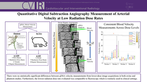
Purpose: Quantitative digital subtraction angiography (qDSA) has been proposed to quantify blood velocity for monitoring treatment progress during blood flow altering interventions. The method requires high frame rate imaging [~ 30 frame per second (fps)] to capture temporal dynamics. This work investigates performance of qDSA in low radiation dose acquisitions to facilitate clinical translation.
Materials and methods: Velocity quantification accuracy was evaluated at five radiation dose rates in vitro and in vivo. Angiographic technique ranged from 30 fps digital subtraction angiography ( at the interventional reference point) down to a 30 fps protocol at 23% higher radiation dose per frame than fluoroscopy ( ). The in vitro setup consisted of a 3D-printed model of a swine hepatic arterial tree connected to a pulsatile displacement pump. Five different flow rates (3.5-8.8 mL/s) were investigated in vitro. Angiography-based fluid velocity measurements were compared across dose rates using ANOVA and Bland-Altman analysis. The experiment was then repeated in a swine study (n = 4).
Results: Radiation dose rate reductions for the lowest dose protocol were 99% and 96% for the phantom and swine study, respectively. No significant difference was found between angiography-based velocity measurements at different dose rates in vitro or in vivo. Bland-Altman analysis found little bias for all lower-dose protocols (range: [- 0.1, 0.1] cm/s), with the widest limits of agreement ([- 3.3, 3.5] cm/s) occurring at the lowest dose protocol.
Conclusions: This study demonstrates the feasibility of quantitative blood velocity measurements from angiographic images acquired at reduced radiation dose rates.
期刊介绍:
CardioVascular and Interventional Radiology (CVIR) is the official journal of the Cardiovascular and Interventional Radiological Society of Europe, and is also the official organ of a number of additional distinguished national and international interventional radiological societies. CVIR publishes double blinded peer-reviewed original research work including clinical and laboratory investigations, technical notes, case reports, works in progress, and letters to the editor, as well as review articles, pictorial essays, editorials, and special invited submissions in the field of vascular and interventional radiology. Beside the communication of the latest research results in this field, it is also the aim of CVIR to support continuous medical education. Articles that are accepted for publication are done so with the understanding that they, or their substantive contents, have not been and will not be submitted to any other publication.
 求助内容:
求助内容: 应助结果提醒方式:


