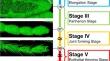
{"title":"Unveiling the cell dynamics during the final shape formation of the tarsus in Drosophila adult leg by live imaging.","authors":"Shotaro Hiraiwa, Shumpei Takeshita, Tensho Terano, Ryuhei Hayashi, Koyo Suzuki, Reiko Tajiri, Tetsuya Kojima","doi":"10.1007/s00427-024-00719-z","DOIUrl":null,"url":null,"abstract":"<p><p>Organisms display a remarkable diversity in their shapes. Although substantial progress has been made in unraveling the mechanisms that govern cell fate determination during development, the mechanisms by which fate-determined cells give rise to the final shapes of organisms remain largely unknown. This study describes in detail the process of the final shape formation of the tarsus, which is near the distal tip of the adult leg, during the pupal stage in Drosophila melanogaster. Days-long live imaging revealed unexpectedly complicated cellular dynamics. The epithelial cells transiently form the intriguing structure, which we named the Parthenon-like structure. The basal surface of the epithelial cells and localization of the basement membrane protein initially show a mesh-like structure and rapidly shrink into the membranous structure during the formation and disappearance of the Parthenon-like structure. Furthermore, macrophage-like cells are observed moving around actively in the Parthenon-like structure and engulfing epithelial cells. The findings in this research are expected to significantly contribute to our understanding of the mechanisms involved in shaping the final structure of the adult tarsus.</p>","PeriodicalId":50588,"journal":{"name":"Development Genes and Evolution","volume":" ","pages":"117-133"},"PeriodicalIF":0.8000,"publicationDate":"2024-12-01","publicationTypes":"Journal Article","fieldsOfStudy":null,"isOpenAccess":false,"openAccessPdf":"https://www.ncbi.nlm.nih.gov/pmc/articles/PMC11611951/pdf/","citationCount":"0","resultStr":null,"platform":"Semanticscholar","paperid":null,"PeriodicalName":"Development Genes and Evolution","FirstCategoryId":"99","ListUrlMain":"https://doi.org/10.1007/s00427-024-00719-z","RegionNum":3,"RegionCategory":"生物学","ArticlePicture":[],"TitleCN":null,"AbstractTextCN":null,"PMCID":null,"EPubDate":"2024/7/8 0:00:00","PubModel":"Epub","JCR":"Q4","JCRName":"CELL BIOLOGY","Score":null,"Total":0}
引用次数: 0
Abstract
Organisms display a remarkable diversity in their shapes. Although substantial progress has been made in unraveling the mechanisms that govern cell fate determination during development, the mechanisms by which fate-determined cells give rise to the final shapes of organisms remain largely unknown. This study describes in detail the process of the final shape formation of the tarsus, which is near the distal tip of the adult leg, during the pupal stage in Drosophila melanogaster. Days-long live imaging revealed unexpectedly complicated cellular dynamics. The epithelial cells transiently form the intriguing structure, which we named the Parthenon-like structure. The basal surface of the epithelial cells and localization of the basement membrane protein initially show a mesh-like structure and rapidly shrink into the membranous structure during the formation and disappearance of the Parthenon-like structure. Furthermore, macrophage-like cells are observed moving around actively in the Parthenon-like structure and engulfing epithelial cells. The findings in this research are expected to significantly contribute to our understanding of the mechanisms involved in shaping the final structure of the adult tarsus.
期刊介绍:
Development Genes and Evolution publishes high-quality reports on all aspects of development biology and evolutionary biology. The journal reports on experimental and bioinformatics work at the systemic, cellular and molecular levels in the field of animal and plant systems, covering key aspects of the following topics:
Embryological and genetic analysis of model and non-model organisms
Genes and pattern formation in invertebrates, vertebrates and plants
Axial patterning, embryonic induction and fate maps
Cellular mechanisms of morphogenesis and organogenesis
Stem cells and regeneration
Functional genomics of developmental processes
Developmental diversity and evolution
Evolution of developmentally relevant genes
Phylogeny of animals and plants
Microevolution
Paleontology.
 求助内容:
求助内容: 应助结果提醒方式:


