{"title":"Subject-Specific Probability Maps of Scalp, Skull and Cerebrospinal Fluid for Cranial Bones Segmentation in Neonatal Cerebral MRIs","authors":"","doi":"10.1016/j.irbm.2024.100844","DOIUrl":null,"url":null,"abstract":"<div><h3>Objectives</h3><p>Segmentation of cranial bones in magnetic resonance images (MRIs) is a challenging and indispensable task to study neonatal brain development and injury. This paper presents a new approach for creating subject-specific probability maps of the scalp, skull and cerebrospinal fluid (CSF) from retrospective bimodal (MR and CT) images acquired from neonates in the gestational age range of 39 to 42 weeks. These maps are subsequently employed for the segmentation of cranial bones in cerebral MRIs from neonates in the same age range.</p></div><div><h3>Material and methods</h3><p>Retrospective MR and CT of neonates with normal head in the gestational age range of 39-42 weeks were preprocessed, segmented semi-automatically and employed as atlas data. For an input MR image acquired from a subject under study, a preprocessing stage and three main processing blocks were performed: First, subject-specific head and intracranial templates and CSF probability map were created using retrospective MR atlas data. Second, the CT atlas data were coregistered to MR templates and the resulted deformation matrices were fed to the next block to create subject-specific scalp and skull probability maps. Finally, some novel performance measures were presented to evaluate the performance of subject-specific CSF, scalp and skull probability maps for skull and intracranial segmentation in neonatal MRIs.</p></div><div><h3>Results</h3><p>The subject-specific probability maps were employed for brain tissue extraction and compared with two public methods such as Brain Extraction Tool (BET) and Brain Surface Extractor (BSE). They were also applied for cranial bone extraction. Then, the similarity in shape between the frontal and occipital sutures (which had been reconstructed from segmented cranial bones) and the ground truth landmarks was evaluated. For this purpose, modified versions of the Dice similarity coefficient (DSC) were used. Finally, a retrospective bimodal (MR-CT) data acquired from a neonate within a short time interval was used for evaluation. After co-alignment of the two images, the DSC and modified Hausdorff distance (MHD) were used to compare the similarity of cranial bones in the MR and CT images.</p></div><div><h3>Conclusion</h3><p>Significant improvements were achieved compared to conventional methods which rely solely on MR image intensities. These advancements hold promise for enhancing neurodevelopmental studies in neonates. The algorithm for creating subject-specific atlases is publicly accessible through a graphical user interface at <span><span>medvispy.ee.kntu.ac.ir</span><svg><path></path></svg></span>.</p></div>","PeriodicalId":14605,"journal":{"name":"Irbm","volume":"45 4","pages":"Article 100844"},"PeriodicalIF":5.6000,"publicationDate":"2024-06-19","publicationTypes":"Journal Article","fieldsOfStudy":null,"isOpenAccess":false,"openAccessPdf":"","citationCount":"0","resultStr":null,"platform":"Semanticscholar","paperid":null,"PeriodicalName":"Irbm","FirstCategoryId":"5","ListUrlMain":"https://www.sciencedirect.com/science/article/pii/S1959031824000253","RegionNum":4,"RegionCategory":"医学","ArticlePicture":[],"TitleCN":null,"AbstractTextCN":null,"PMCID":null,"EPubDate":"","PubModel":"","JCR":"Q1","JCRName":"ENGINEERING, BIOMEDICAL","Score":null,"Total":0}
引用次数: 0
Abstract
Objectives
Segmentation of cranial bones in magnetic resonance images (MRIs) is a challenging and indispensable task to study neonatal brain development and injury. This paper presents a new approach for creating subject-specific probability maps of the scalp, skull and cerebrospinal fluid (CSF) from retrospective bimodal (MR and CT) images acquired from neonates in the gestational age range of 39 to 42 weeks. These maps are subsequently employed for the segmentation of cranial bones in cerebral MRIs from neonates in the same age range.
Material and methods
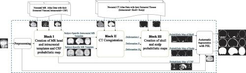
Retrospective MR and CT of neonates with normal head in the gestational age range of 39-42 weeks were preprocessed, segmented semi-automatically and employed as atlas data. For an input MR image acquired from a subject under study, a preprocessing stage and three main processing blocks were performed: First, subject-specific head and intracranial templates and CSF probability map were created using retrospective MR atlas data. Second, the CT atlas data were coregistered to MR templates and the resulted deformation matrices were fed to the next block to create subject-specific scalp and skull probability maps. Finally, some novel performance measures were presented to evaluate the performance of subject-specific CSF, scalp and skull probability maps for skull and intracranial segmentation in neonatal MRIs.
Results
The subject-specific probability maps were employed for brain tissue extraction and compared with two public methods such as Brain Extraction Tool (BET) and Brain Surface Extractor (BSE). They were also applied for cranial bone extraction. Then, the similarity in shape between the frontal and occipital sutures (which had been reconstructed from segmented cranial bones) and the ground truth landmarks was evaluated. For this purpose, modified versions of the Dice similarity coefficient (DSC) were used. Finally, a retrospective bimodal (MR-CT) data acquired from a neonate within a short time interval was used for evaluation. After co-alignment of the two images, the DSC and modified Hausdorff distance (MHD) were used to compare the similarity of cranial bones in the MR and CT images.
Conclusion
Significant improvements were achieved compared to conventional methods which rely solely on MR image intensities. These advancements hold promise for enhancing neurodevelopmental studies in neonates. The algorithm for creating subject-specific atlases is publicly accessible through a graphical user interface at medvispy.ee.kntu.ac.ir.
期刊介绍:
IRBM is the journal of the AGBM (Alliance for engineering in Biology an Medicine / Alliance pour le génie biologique et médical) and the SFGBM (BioMedical Engineering French Society / Société française de génie biologique médical) and the AFIB (French Association of Biomedical Engineers / Association française des ingénieurs biomédicaux).
As a vehicle of information and knowledge in the field of biomedical technologies, IRBM is devoted to fundamental as well as clinical research. Biomedical engineering and use of new technologies are the cornerstones of IRBM, providing authors and users with the latest information. Its six issues per year propose reviews (state-of-the-art and current knowledge), original articles directed at fundamental research and articles focusing on biomedical engineering. All articles are submitted to peer reviewers acting as guarantors for IRBM''s scientific and medical content. The field covered by IRBM includes all the discipline of Biomedical engineering. Thereby, the type of papers published include those that cover the technological and methodological development in:
-Physiological and Biological Signal processing (EEG, MEG, ECG…)-
Medical Image processing-
Biomechanics-
Biomaterials-
Medical Physics-
Biophysics-
Physiological and Biological Sensors-
Information technologies in healthcare-
Disability research-
Computational physiology-
…
 求助内容:
求助内容: 应助结果提醒方式:


