{"title":"Silibinin promotes healing in spinal cord injury through anti-ferroptotic mechanisms","authors":"Arman Vahabi, Anıl Murat Öztürk, Bünyamin Kılıçlı, Derviş Birim, Gizem Kaftan Öcal, Taner Dağcı, Güliz Armağan","doi":"10.1002/jsp2.1344","DOIUrl":null,"url":null,"abstract":"<div>\n \n \n <section>\n \n <h3> Study Design</h3>\n \n <p>Pre-clinical animal experiment.</p>\n </section>\n \n <section>\n \n <h3> Objective</h3>\n \n <p>In this study, we investigated therapeutic effects of silibinin in a spinal cord injury (SCI) model. In SCI, loss of cells due to secondary damage mechanisms exceeds that caused by primary damage. Ferroptosis, which is iron-dependent non-apoptotic cell death, is shown to be influential in the pathogenesis of SCI.</p>\n </section>\n \n <section>\n \n <h3> Methods</h3>\n \n <p>The study was conducted as an in vivo experiment using a total of 78 adult male/female Sprague Dawley rats. Groups were as follows: Sham, SCI, deferoxamine (DFO) treatment, and silibinin treatment. There were subgroups with follow-up periods of 24 h, 72 h, and 6 weeks in all groups. Malondialdehyde (MDA), glutathione (GSH), and Fe<sup>2+</sup> levels were measured by spectrophotometry. Glutathione peroxidase-4 (GPX4), ferroportin (FPN), transferrin receptor (TfR1), and 4-hydroxynonenal (4-HNE)-modified protein levels were assessed by Western blotting. Functional recovery was assessed using Basso–Beattie–Bresnahan test.</p>\n </section>\n \n <section>\n \n <h3> Results</h3>\n \n <p>Silibinin achieved significant suppression in MDA and 4-HNE levels compared to the SCI both in 72-h and 6 weeks group (<i>p</i> < 0.05). GSH, GPX4, and FNP levels were found to be significantly higher in the silibinin 24 h, 72 h, and 6 weeks group compared to corresponding SCI groups (<i>p</i> < 0.05). Significant reduction in iron levels was observed in silibinin treated rats in 72 h and 6 weeks group (<i>p</i> < 0.05). Silibinin substantially suppressed TfR1 levels in 24 h and 72 h groups (<i>p</i> < 0.05). Significant difference among recovery capacities was observed as follows: Silibinin > DFO > SCI (<i>p</i> < 0.05).</p>\n </section>\n \n <section>\n \n <h3> Conclusion</h3>\n \n <p>Impact of silibinin on iron metabolism and lipid peroxidation, both of which are features of ferroptosis, may contribute to therapeutic activity. Within this context, our findings posit silibinin as a potential therapeutic candidate possessing antiferroptotic properties in SCI model. Therapeutic agents capable of effectively and safely mitigating ferroptotic cell death hold the potential to be critical points of future clinical investigations.</p>\n </section>\n </div>","PeriodicalId":14876,"journal":{"name":"JOR Spine","volume":"7 3","pages":""},"PeriodicalIF":3.4000,"publicationDate":"2024-07-01","publicationTypes":"Journal Article","fieldsOfStudy":null,"isOpenAccess":false,"openAccessPdf":"https://www.ncbi.nlm.nih.gov/pmc/articles/PMC11217020/pdf/","citationCount":"0","resultStr":null,"platform":"Semanticscholar","paperid":null,"PeriodicalName":"JOR Spine","FirstCategoryId":"3","ListUrlMain":"https://onlinelibrary.wiley.com/doi/10.1002/jsp2.1344","RegionNum":3,"RegionCategory":"医学","ArticlePicture":[],"TitleCN":null,"AbstractTextCN":null,"PMCID":null,"EPubDate":"","PubModel":"","JCR":"Q1","JCRName":"ORTHOPEDICS","Score":null,"Total":0}
引用次数: 0
Abstract
Study Design
Pre-clinical animal experiment.
Objective
In this study, we investigated therapeutic effects of silibinin in a spinal cord injury (SCI) model. In SCI, loss of cells due to secondary damage mechanisms exceeds that caused by primary damage. Ferroptosis, which is iron-dependent non-apoptotic cell death, is shown to be influential in the pathogenesis of SCI.
Methods
The study was conducted as an in vivo experiment using a total of 78 adult male/female Sprague Dawley rats. Groups were as follows: Sham, SCI, deferoxamine (DFO) treatment, and silibinin treatment. There were subgroups with follow-up periods of 24 h, 72 h, and 6 weeks in all groups. Malondialdehyde (MDA), glutathione (GSH), and Fe2+ levels were measured by spectrophotometry. Glutathione peroxidase-4 (GPX4), ferroportin (FPN), transferrin receptor (TfR1), and 4-hydroxynonenal (4-HNE)-modified protein levels were assessed by Western blotting. Functional recovery was assessed using Basso–Beattie–Bresnahan test.
Results
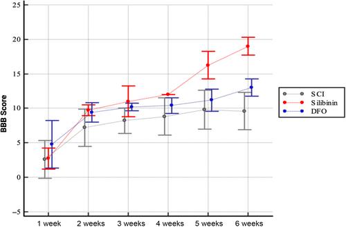
Silibinin achieved significant suppression in MDA and 4-HNE levels compared to the SCI both in 72-h and 6 weeks group (p < 0.05). GSH, GPX4, and FNP levels were found to be significantly higher in the silibinin 24 h, 72 h, and 6 weeks group compared to corresponding SCI groups (p < 0.05). Significant reduction in iron levels was observed in silibinin treated rats in 72 h and 6 weeks group (p < 0.05). Silibinin substantially suppressed TfR1 levels in 24 h and 72 h groups (p < 0.05). Significant difference among recovery capacities was observed as follows: Silibinin > DFO > SCI (p < 0.05).
Conclusion
Impact of silibinin on iron metabolism and lipid peroxidation, both of which are features of ferroptosis, may contribute to therapeutic activity. Within this context, our findings posit silibinin as a potential therapeutic candidate possessing antiferroptotic properties in SCI model. Therapeutic agents capable of effectively and safely mitigating ferroptotic cell death hold the potential to be critical points of future clinical investigations.