Nargiz Ahmadova, Mustafa Kayabaşı, Seher Köksaldı, Eda Hümaz, Ali Osman Saatci
{"title":"Multimodal Imaging Characteristics and Diagnostic Value of Choroidal Nodules in Patients with Neurofibromatosis Type 1.","authors":"Nargiz Ahmadova, Mustafa Kayabaşı, Seher Köksaldı, Eda Hümaz, Ali Osman Saatci","doi":"10.4274/tjo.galenos.2024.48017","DOIUrl":null,"url":null,"abstract":"<p><strong>Objectives: </strong>Yasunari nodules are choroidal lesions observed in patients diagnosed with neurofibromatosis type 1 (NF-1) and characterized by relatively irregular dome-shaped, plaque-like, or patchy boundaries. The present study examines the multimodal imaging characteristics of Yasunari nodules and their value in the diagnosis of NF-1.</p><p><strong>Materials and methods: </strong>Medical records including optical coherence tomography (OCT), enhanced depth imaging OCT, infrared reflectance (IR) imaging, OCT angiography, and color fundus images of NF-1 patients who were examined at the Department of Ophthalmology in Dokuz Eylül University Faculty of Medicine between January 2022 and December 2023 were retrospectively reviewed for the presence of Yasunari nodules.</p><p><strong>Results: </strong>A total of 54 eyes of 27 patients were included in the study. At least one choroidal nodule was detected on IR imaging in 52 eyes (96.3%). In 31 (72.1%) of the 43 eyes (79.6%) with available high-quality OCT angiography images, choroidal nodules were observed as areas showing a flow deficit in the choriocapillaris layer. Of the total 54 eyes included, Lisch nodules without choroidal nodules were observed in 2 eyes (3.7%). In 16 eyes (29.6%), Lisch nodules were not detected despite the presence of choroidal nodules. Both Lisch nodules and choroidal nodules were detected in the other 36 eyes (66.7%).</p><p><strong>Conclusion: </strong>Yasunari nodules are frequently observed in NF-1 cases and can be easily detected with multimodal imaging techniques, especially IR imaging. The ability to visualize choroidal nodules before the appearance of Lisch nodules demonstrates the importance of Yasunari nodules in the diagnosis of NF-1.</p>","PeriodicalId":23373,"journal":{"name":"Turkish Journal of Ophthalmology","volume":"54 3","pages":"140-148"},"PeriodicalIF":0.0000,"publicationDate":"2024-06-28","publicationTypes":"Journal Article","fieldsOfStudy":null,"isOpenAccess":false,"openAccessPdf":"https://www.ncbi.nlm.nih.gov/pmc/articles/PMC11589307/pdf/","citationCount":"0","resultStr":null,"platform":"Semanticscholar","paperid":null,"PeriodicalName":"Turkish Journal of Ophthalmology","FirstCategoryId":"1085","ListUrlMain":"https://doi.org/10.4274/tjo.galenos.2024.48017","RegionNum":0,"RegionCategory":null,"ArticlePicture":[],"TitleCN":null,"AbstractTextCN":null,"PMCID":null,"EPubDate":"","PubModel":"","JCR":"Q3","JCRName":"Medicine","Score":null,"Total":0}
引用次数: 0
Abstract
Objectives: Yasunari nodules are choroidal lesions observed in patients diagnosed with neurofibromatosis type 1 (NF-1) and characterized by relatively irregular dome-shaped, plaque-like, or patchy boundaries. The present study examines the multimodal imaging characteristics of Yasunari nodules and their value in the diagnosis of NF-1.
Materials and methods: Medical records including optical coherence tomography (OCT), enhanced depth imaging OCT, infrared reflectance (IR) imaging, OCT angiography, and color fundus images of NF-1 patients who were examined at the Department of Ophthalmology in Dokuz Eylül University Faculty of Medicine between January 2022 and December 2023 were retrospectively reviewed for the presence of Yasunari nodules.
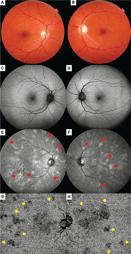
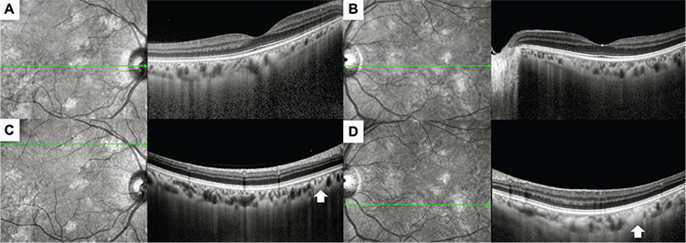
Results: A total of 54 eyes of 27 patients were included in the study. At least one choroidal nodule was detected on IR imaging in 52 eyes (96.3%). In 31 (72.1%) of the 43 eyes (79.6%) with available high-quality OCT angiography images, choroidal nodules were observed as areas showing a flow deficit in the choriocapillaris layer. Of the total 54 eyes included, Lisch nodules without choroidal nodules were observed in 2 eyes (3.7%). In 16 eyes (29.6%), Lisch nodules were not detected despite the presence of choroidal nodules. Both Lisch nodules and choroidal nodules were detected in the other 36 eyes (66.7%).
Conclusion: Yasunari nodules are frequently observed in NF-1 cases and can be easily detected with multimodal imaging techniques, especially IR imaging. The ability to visualize choroidal nodules before the appearance of Lisch nodules demonstrates the importance of Yasunari nodules in the diagnosis of NF-1.
期刊介绍:
The Turkish Journal of Ophthalmology (TJO) is the only scientific periodical publication of the Turkish Ophthalmological Association and has been published since January 1929. In its early years, the journal was published in Turkish and French. Although there were temporary interruptions in the publication of the journal due to various challenges, the Turkish Journal of Ophthalmology has been published continually from 1971 to the present. The target audience includes specialists and physicians in training in ophthalmology in all relevant disciplines.


 求助内容:
求助内容: 应助结果提醒方式:


