Kayvan Samimi, Ojaswi Pasachhe, Emmanuel Contreras Guzman, Jeremiah Riendeau, Amani A. Gillette, Dan L. Pham, Kasia J. Wiech, Darcie L. Moore, Melissa C. Skala
{"title":"Autofluorescence lifetime flow cytometry with time-correlated single photon counting","authors":"Kayvan Samimi, Ojaswi Pasachhe, Emmanuel Contreras Guzman, Jeremiah Riendeau, Amani A. Gillette, Dan L. Pham, Kasia J. Wiech, Darcie L. Moore, Melissa C. Skala","doi":"10.1002/cyto.a.24883","DOIUrl":null,"url":null,"abstract":"<p>Autofluorescence lifetime imaging microscopy (FLIM) is sensitive to metabolic changes in single cells based on changes in the protein-binding activities of the metabolic co-enzymes NAD(P)H. However, FLIM typically relies on time-correlated single-photon counting (TCSPC) detection electronics on laser-scanning microscopes, which are expensive, low-throughput, and require substantial post-processing time for cell segmentation and analysis. Here, we present a fluorescence lifetime-sensitive flow cytometer that offers the same TCSPC temporal resolution in a flow geometry, with low-cost single-photon excitation sources, a throughput of tens of cells per second, and real-time single-cell analysis. The system uses a 375 nm picosecond-pulsed diode laser operating at 50 MHz, alkali photomultiplier tubes, an FPGA-based time tagger, and can provide real-time phasor-based classification (i.e., gating) of flowing cells. A CMOS camera produces simultaneous brightfield images using far-red illumination. A second PMT provides two-color analysis. Cells are injected into the microfluidic channel using a syringe pump at 2–5 mm/s with nearly 5 ms integration time per cell, resulting in a light dose of 2.65 J/cm<sup>2</sup> that is well below damage thresholds (25 J/cm<sup>2</sup> at 375 nm). Our results show that cells remain viable after measurement, and the system is sensitive to autofluorescence lifetime changes in Jurkat T cells with metabolic perturbation (sodium cyanide), quiescent versus activated (CD3/CD28/CD2) primary human T cells, and quiescent versus activated primary adult mouse neural stem cells, consistent with prior studies using multiphoton FLIM. This TCSPC-based autofluorescence lifetime flow cytometer provides a valuable label-free method for real-time analysis of single-cell function and metabolism with higher throughput than laser-scanning microscopy systems.</p>","PeriodicalId":11068,"journal":{"name":"Cytometry Part A","volume":"105 8","pages":"607-620"},"PeriodicalIF":2.1000,"publicationDate":"2024-06-28","publicationTypes":"Journal Article","fieldsOfStudy":null,"isOpenAccess":false,"openAccessPdf":"https://onlinelibrary.wiley.com/doi/epdf/10.1002/cyto.a.24883","citationCount":"0","resultStr":null,"platform":"Semanticscholar","paperid":null,"PeriodicalName":"Cytometry Part A","FirstCategoryId":"99","ListUrlMain":"https://onlinelibrary.wiley.com/doi/10.1002/cyto.a.24883","RegionNum":4,"RegionCategory":"生物学","ArticlePicture":[],"TitleCN":null,"AbstractTextCN":null,"PMCID":null,"EPubDate":"","PubModel":"","JCR":"Q3","JCRName":"BIOCHEMICAL RESEARCH METHODS","Score":null,"Total":0}
引用次数: 0
Abstract
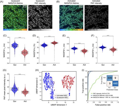
Autofluorescence lifetime imaging microscopy (FLIM) is sensitive to metabolic changes in single cells based on changes in the protein-binding activities of the metabolic co-enzymes NAD(P)H. However, FLIM typically relies on time-correlated single-photon counting (TCSPC) detection electronics on laser-scanning microscopes, which are expensive, low-throughput, and require substantial post-processing time for cell segmentation and analysis. Here, we present a fluorescence lifetime-sensitive flow cytometer that offers the same TCSPC temporal resolution in a flow geometry, with low-cost single-photon excitation sources, a throughput of tens of cells per second, and real-time single-cell analysis. The system uses a 375 nm picosecond-pulsed diode laser operating at 50 MHz, alkali photomultiplier tubes, an FPGA-based time tagger, and can provide real-time phasor-based classification (i.e., gating) of flowing cells. A CMOS camera produces simultaneous brightfield images using far-red illumination. A second PMT provides two-color analysis. Cells are injected into the microfluidic channel using a syringe pump at 2–5 mm/s with nearly 5 ms integration time per cell, resulting in a light dose of 2.65 J/cm2 that is well below damage thresholds (25 J/cm2 at 375 nm). Our results show that cells remain viable after measurement, and the system is sensitive to autofluorescence lifetime changes in Jurkat T cells with metabolic perturbation (sodium cyanide), quiescent versus activated (CD3/CD28/CD2) primary human T cells, and quiescent versus activated primary adult mouse neural stem cells, consistent with prior studies using multiphoton FLIM. This TCSPC-based autofluorescence lifetime flow cytometer provides a valuable label-free method for real-time analysis of single-cell function and metabolism with higher throughput than laser-scanning microscopy systems.
期刊介绍:
Cytometry Part A, the journal of quantitative single-cell analysis, features original research reports and reviews of innovative scientific studies employing quantitative single-cell measurement, separation, manipulation, and modeling techniques, as well as original articles on mechanisms of molecular and cellular functions obtained by cytometry techniques.
The journal welcomes submissions from multiple research fields that fully embrace the study of the cytome:
Biomedical Instrumentation Engineering
Biophotonics
Bioinformatics
Cell Biology
Computational Biology
Data Science
Immunology
Parasitology
Microbiology
Neuroscience
Cancer
Stem Cells
Tissue Regeneration.
 求助内容:
求助内容: 应助结果提醒方式:


