{"title":"Muscle resection biopsy during peroral endoscopic myotomy in a patient with achalasia","authors":"Shinya Hoki, Hirofumi Abe, Chise Ueda","doi":"10.1111/den.14871","DOIUrl":null,"url":null,"abstract":"<p>Esophageal achalasia is an esophageal motility disorder, primarily characterized by degeneration of the esophageal myenteric plexus.<span><sup>1</sup></span> Although the myenteric plexus is the primary concern during pathogenesis of achalasia,<span><sup>2</sup></span> a method for endoscopically sampling it has not yet been established. We report a novel sampling method—muscle resection biopsy—designed to sample the myenteric plexus while distinguishing between the circular and longitudinal muscle layers during peroral endoscopic myotomy (Video S1). A submucosal tunnel was first created and a small full-thickness muscle incision was made just above the lower esophageal sphincter using a needle-type knife (FlushKnife BTS3.0; FUJIFILM Holdings Corporation, Tokyo, Japan) and laterally extended to both sides, forming a U shape (Fig. 1a). A hemostatic clip (EZclip; Olympus Corporation, Tokyo, Japan), with one arm marked in red, was applied to the shaped muscle layers with the marked arm on the luminal side (Fig. 1b,c). We then excised the remaining muscle layers using a snare (SD-221L-25; Olympus Corporation; Fig. 1d) and collected the resected tissue.</p><p>Peroral endoscopic muscle biopsy using a submucosal tunnel has been recognized as a simple and useful sampling method for evaluating eosinophilic infiltration and fibrosis in the muscle layer.<span><sup>3</sup></span> However, the small size of biopsy samples and tissue damage caused by biopsy forceps make identifying the myenteric plexus and preserving the structures of both the circular and longitudinal muscle layers challenging. Although this method is time-consuming, requires skillful manipulation of an endoknife, and has potential risk of bleeding, it enables the collection of large, undamaged, tissues via biopsy forceps and allows for identification of the myenteric plexus and muscle layers while preserving the microscopic structure of the luminal wall (Fig. 2). Histopathological information obtained by this approach can be useful for assessing the microenvironment underlying the neurodegeneration in combination with immunohistochemical staining results.</p><p>Authors declare no conflict of interest for this article.</p>","PeriodicalId":159,"journal":{"name":"Digestive Endoscopy","volume":"36 9","pages":"1052-1053"},"PeriodicalIF":5.0000,"publicationDate":"2024-06-24","publicationTypes":"Journal Article","fieldsOfStudy":null,"isOpenAccess":false,"openAccessPdf":"https://onlinelibrary.wiley.com/doi/epdf/10.1111/den.14871","citationCount":"0","resultStr":null,"platform":"Semanticscholar","paperid":null,"PeriodicalName":"Digestive Endoscopy","FirstCategoryId":"3","ListUrlMain":"https://onlinelibrary.wiley.com/doi/10.1111/den.14871","RegionNum":2,"RegionCategory":"医学","ArticlePicture":[],"TitleCN":null,"AbstractTextCN":null,"PMCID":null,"EPubDate":"","PubModel":"","JCR":"Q1","JCRName":"GASTROENTEROLOGY & HEPATOLOGY","Score":null,"Total":0}
引用次数: 0
Abstract
Esophageal achalasia is an esophageal motility disorder, primarily characterized by degeneration of the esophageal myenteric plexus.1 Although the myenteric plexus is the primary concern during pathogenesis of achalasia,2 a method for endoscopically sampling it has not yet been established. We report a novel sampling method—muscle resection biopsy—designed to sample the myenteric plexus while distinguishing between the circular and longitudinal muscle layers during peroral endoscopic myotomy (Video S1). A submucosal tunnel was first created and a small full-thickness muscle incision was made just above the lower esophageal sphincter using a needle-type knife (FlushKnife BTS3.0; FUJIFILM Holdings Corporation, Tokyo, Japan) and laterally extended to both sides, forming a U shape (Fig. 1a). A hemostatic clip (EZclip; Olympus Corporation, Tokyo, Japan), with one arm marked in red, was applied to the shaped muscle layers with the marked arm on the luminal side (Fig. 1b,c). We then excised the remaining muscle layers using a snare (SD-221L-25; Olympus Corporation; Fig. 1d) and collected the resected tissue.
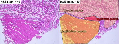
Peroral endoscopic muscle biopsy using a submucosal tunnel has been recognized as a simple and useful sampling method for evaluating eosinophilic infiltration and fibrosis in the muscle layer.3 However, the small size of biopsy samples and tissue damage caused by biopsy forceps make identifying the myenteric plexus and preserving the structures of both the circular and longitudinal muscle layers challenging. Although this method is time-consuming, requires skillful manipulation of an endoknife, and has potential risk of bleeding, it enables the collection of large, undamaged, tissues via biopsy forceps and allows for identification of the myenteric plexus and muscle layers while preserving the microscopic structure of the luminal wall (Fig. 2). Histopathological information obtained by this approach can be useful for assessing the microenvironment underlying the neurodegeneration in combination with immunohistochemical staining results.
Authors declare no conflict of interest for this article.
期刊介绍:
Digestive Endoscopy (DEN) is the official journal of the Japan Gastroenterological Endoscopy Society, the Asian Pacific Society for Digestive Endoscopy and the World Endoscopy Organization. Digestive Endoscopy serves as a medium for presenting original articles that offer significant contributions to knowledge in the broad field of endoscopy. The Journal also includes Reviews, Original Articles, How I Do It, Case Reports (only of exceptional interest and novelty are accepted), Letters, Techniques and Images, abstracts and news items that may be of interest to endoscopists.

 求助内容:
求助内容: 应助结果提醒方式:


