Development and validation of a smartphone-based deep-learning-enabled system to detect middle-ear conditions in otoscopic images
IF 12.4
1区 医学
Q1 HEALTH CARE SCIENCES & SERVICES
引用次数: 0
Abstract
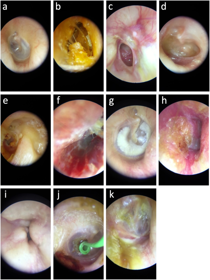
Middle-ear conditions are common causes of primary care visits, hearing impairment, and inappropriate antibiotic use. Deep learning (DL) may assist clinicians in interpreting otoscopic images. This study included patients over 5 years old from an ambulatory ENT practice in Strasbourg, France, between 2013 and 2020. Digital otoscopic images were obtained using a smartphone-attached otoscope (Smart Scope, Karl Storz, Germany) and labeled by a senior ENT specialist across 11 diagnostic classes (reference standard). An Inception-v2 DL model was trained using 41,664 otoscopic images, and its diagnostic accuracy was evaluated by calculating class-specific estimates of sensitivity and specificity. The model was then incorporated into a smartphone app called i-Nside. The DL model was evaluated on a validation set of 3,962 images and a held-out test set comprising 326 images. On the validation set, all class-specific estimates of sensitivity and specificity exceeded 98%. On the test set, the DL model achieved a sensitivity of 99.0% (95% confidence interval: 94.5–100) and a specificity of 95.2% (91.5–97.6) for the binary classification of normal vs. abnormal images; wax plugs were detected with a sensitivity of 100% (94.6–100) and specificity of 97.7% (95.0–99.1); other class-specific estimates of sensitivity and specificity ranged from 33.3% to 92.3% and 96.0% to 100%, respectively. We present an end-to-end DL-enabled system able to achieve expert-level diagnostic accuracy for identifying normal tympanic aspects and wax plugs within digital otoscopic images. However, the system’s performance varied for other middle-ear conditions. Further prospective validation is necessary before wider clinical deployment.


开发和验证基于智能手机的深度学习系统,以检测耳镜图像中的中耳状况。
中耳疾病是导致初级保健就诊、听力受损和抗生素使用不当的常见原因。深度学习(DL)可帮助临床医生解读耳镜图像。这项研究纳入了法国斯特拉斯堡一家门诊耳鼻喉科诊所在2013年至2020年间收治的5岁以上患者。数字耳镜图像通过智能手机连接的耳镜(Smart Scope,德国卡尔-斯托尔兹公司)获得,并由资深耳鼻喉科专家按照 11 个诊断类别(参考标准)进行标注。使用 41,664 张耳镜图像对 Inception-v2 DL 模型进行了训练,并通过计算特定类别的灵敏度和特异性估计值来评估其诊断准确性。随后,该模型被整合到一款名为 i-Nside 的智能手机应用程序中。DL 模型在由 3962 张图像组成的验证集和 326 张图像组成的保留测试集上进行了评估。在验证集上,所有类别的灵敏度和特异性估计值都超过了 98%。在测试集上,DL 模型对正常与异常图像进行二元分类的灵敏度为 99.0%(95% 置信区间:94.5-100),特异性为 95.2%(91.5-97.6);检测到蜡栓的灵敏度为 100%(94.6-100),特异性为 97.7%(95.0-99.1);其他类别的灵敏度和特异性估计值分别为 33.3% 到 92.3%,96.0% 到 100%。我们介绍的端到端 DL 支持系统能够达到专家级诊断准确度,在数字耳镜图像中识别正常鼓室和蜡栓。然而,该系统对其他中耳病症的表现却不尽相同。在更广泛的临床应用之前,有必要进行进一步的前瞻性验证。
本文章由计算机程序翻译,如有差异,请以英文原文为准。
求助全文
约1分钟内获得全文
求助全文
来源期刊

NPJ Digital Medicine
Multiple-
CiteScore
25.10
自引率
3.30%
发文量
170
审稿时长
15 weeks
期刊介绍:
npj Digital Medicine is an online open-access journal that focuses on publishing peer-reviewed research in the field of digital medicine. The journal covers various aspects of digital medicine, including the application and implementation of digital and mobile technologies in clinical settings, virtual healthcare, and the use of artificial intelligence and informatics.
The primary goal of the journal is to support innovation and the advancement of healthcare through the integration of new digital and mobile technologies. When determining if a manuscript is suitable for publication, the journal considers four important criteria: novelty, clinical relevance, scientific rigor, and digital innovation.
 求助内容:
求助内容: 应助结果提醒方式:


