{"title":"Cerebral air embolism followed by endoscopic balloon dilatation for esophageal strictures","authors":"Ryoichi Shoji, Naruaki Otake, Takeo Nagura, Jushi Numata, Junya Tsurukiri","doi":"10.1002/ams2.971","DOIUrl":null,"url":null,"abstract":"<p>A 59-year-old male patient underwent esophageal endoscopic balloon dilatation (BD) under sedation for esophageal strictures at another hospital. He suddenly became unresponsive post-procedure, resulting in coma and undergoing mechanical ventilation. Head computed tomography (CT) revealed multiple air emboli within the parenchyma, and cerebral air embolism (CAE) was diagnosed. Additionally, abdominal CT revealed air in the portal venous system. Diffusion-weighted magnetic resonance imaging after 2 days confirmed areas of acute ischemia involving the right frontal, temporal, and occipital lobes (Figure 1). Microbubble-enhanced echocardiography revealed no evidence of a cardiac shunt. His consciousness gradually improved over several days without undergoing hyperbaric oxygen therapy, and he was discharged for rehabilitation in left hemiplegia after 30 days.</p><p>CAE is a rare, potentially catastrophic iatrogenic complication.<span><sup>1</sup></span> Four reports on esophageal endoscopic BD-induced CAE were retrieved from PubMed, but the pathogenesis without obtaining whole-body imaging remains unclear.<span><sup>2-5</sup></span> The venous air embolism, including the portal vein and the cerebrum, explained the present case. This is the first report on the pathogenesis visualized as supporting evidence of CAE during endoscopic procedures. The mechanism includes air transgression from the esophageal mucosa to the vasculature and flowing into the portal venous system through portal–esophageal vein radicles transected during the procedure.</p><p>All authors declare that they have no conflict of interest, and the manuscript has not been previously published; the manuscript is not under consideration for publication elsewhere.</p><p>Approval of the research protocol: N/A.</p><p>Informed consent: Written informed consent was obtained from the patient for publication of this case report and accompanying images.</p><p>Registry and the registration no. of the study/trial: N/A.</p><p>Animal studies: N/A.</p>","PeriodicalId":7196,"journal":{"name":"Acute Medicine & Surgery","volume":"11 1","pages":""},"PeriodicalIF":1.3000,"publicationDate":"2024-06-14","publicationTypes":"Journal Article","fieldsOfStudy":null,"isOpenAccess":false,"openAccessPdf":"https://onlinelibrary.wiley.com/doi/epdf/10.1002/ams2.971","citationCount":"0","resultStr":null,"platform":"Semanticscholar","paperid":null,"PeriodicalName":"Acute Medicine & Surgery","FirstCategoryId":"1085","ListUrlMain":"https://onlinelibrary.wiley.com/doi/10.1002/ams2.971","RegionNum":0,"RegionCategory":null,"ArticlePicture":[],"TitleCN":null,"AbstractTextCN":null,"PMCID":null,"EPubDate":"","PubModel":"","JCR":"Q2","JCRName":"MEDICINE, GENERAL & INTERNAL","Score":null,"Total":0}
引用次数: 0
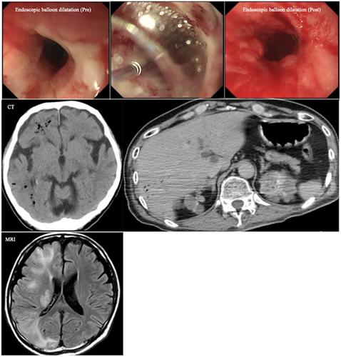
Abstract
A 59-year-old male patient underwent esophageal endoscopic balloon dilatation (BD) under sedation for esophageal strictures at another hospital. He suddenly became unresponsive post-procedure, resulting in coma and undergoing mechanical ventilation. Head computed tomography (CT) revealed multiple air emboli within the parenchyma, and cerebral air embolism (CAE) was diagnosed. Additionally, abdominal CT revealed air in the portal venous system. Diffusion-weighted magnetic resonance imaging after 2 days confirmed areas of acute ischemia involving the right frontal, temporal, and occipital lobes (Figure 1). Microbubble-enhanced echocardiography revealed no evidence of a cardiac shunt. His consciousness gradually improved over several days without undergoing hyperbaric oxygen therapy, and he was discharged for rehabilitation in left hemiplegia after 30 days.
CAE is a rare, potentially catastrophic iatrogenic complication.1 Four reports on esophageal endoscopic BD-induced CAE were retrieved from PubMed, but the pathogenesis without obtaining whole-body imaging remains unclear.2-5 The venous air embolism, including the portal vein and the cerebrum, explained the present case. This is the first report on the pathogenesis visualized as supporting evidence of CAE during endoscopic procedures. The mechanism includes air transgression from the esophageal mucosa to the vasculature and flowing into the portal venous system through portal–esophageal vein radicles transected during the procedure.
All authors declare that they have no conflict of interest, and the manuscript has not been previously published; the manuscript is not under consideration for publication elsewhere.
Approval of the research protocol: N/A.
Informed consent: Written informed consent was obtained from the patient for publication of this case report and accompanying images.
Registry and the registration no. of the study/trial: N/A.

 求助内容:
求助内容: 应助结果提醒方式:


