Nicholas Chen Yi Png, Winfred Xi Tai Goh, Clement Wenhao Chan
{"title":"Metaplastic breast cancer masquerading as a recurrent haematoma: A case report.","authors":"Nicholas Chen Yi Png, Winfred Xi Tai Goh, Clement Wenhao Chan","doi":"10.3233/BD-240006","DOIUrl":null,"url":null,"abstract":"<p><p>An 85-year-old Chinese lady presented with a 5-day history of a painless left breast lump. There was no fever, nipple discharge, or history of trauma. She had a past medical history of atrial fibrillation that was managed with an oral anticoagulant. Mammography demonstrated a dense mass in the upper outer quadrant of the left breast. Ultrasound showed an irregular, heterogeneous 4.7 cm lesion containing debris and cystic spaces with raised peripheral vascularity at the 2 o'clock position, 3 cm from nipple. No internal vascularity was detected. This was managed as a haematoma and rivaroxaban was withheld. Follow-up imaging 3-weeks later showed persistence of the lesion. Bedside needle aspiration yielded haemoserous fluid with immediate reduction in size of the lesion. However, 2 weeks after aspiration, there was recurrence of the 'haematoma'. Multidisciplinary review of the clinical history, examination and imaging was sought, and biopsy of the irregularly thickened areas with vascularity along the periphery of the lesion was recommended. Vacuum-assisted biopsy was performed, and histology returned as metaplastic carcinoma. A recurring 'haematoma' should always prompt a search for a secondary cause, with features such as irregular thickened walls and papillary/nodular components requiring further evaluation with biopsy for histopathological correlation.</p>","PeriodicalId":9224,"journal":{"name":"Breast disease","volume":"43 1","pages":"187-191"},"PeriodicalIF":0.0000,"publicationDate":"2024-01-01","publicationTypes":"Journal Article","fieldsOfStudy":null,"isOpenAccess":false,"openAccessPdf":"https://www.ncbi.nlm.nih.gov/pmc/articles/PMC11191430/pdf/","citationCount":"0","resultStr":null,"platform":"Semanticscholar","paperid":null,"PeriodicalName":"Breast disease","FirstCategoryId":"1085","ListUrlMain":"https://doi.org/10.3233/BD-240006","RegionNum":0,"RegionCategory":null,"ArticlePicture":[],"TitleCN":null,"AbstractTextCN":null,"PMCID":null,"EPubDate":"","PubModel":"","JCR":"","JCRName":"","Score":null,"Total":0}
引用次数: 0
Abstract
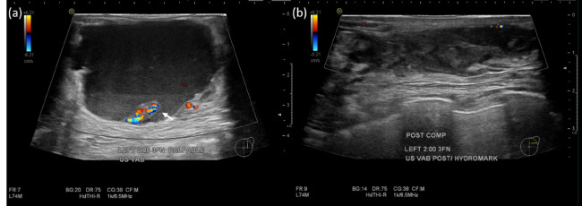
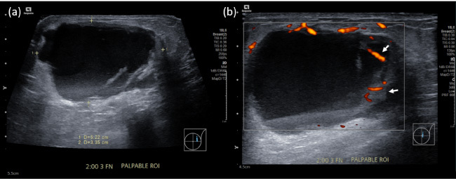
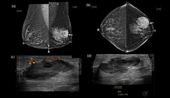
An 85-year-old Chinese lady presented with a 5-day history of a painless left breast lump. There was no fever, nipple discharge, or history of trauma. She had a past medical history of atrial fibrillation that was managed with an oral anticoagulant. Mammography demonstrated a dense mass in the upper outer quadrant of the left breast. Ultrasound showed an irregular, heterogeneous 4.7 cm lesion containing debris and cystic spaces with raised peripheral vascularity at the 2 o'clock position, 3 cm from nipple. No internal vascularity was detected. This was managed as a haematoma and rivaroxaban was withheld. Follow-up imaging 3-weeks later showed persistence of the lesion. Bedside needle aspiration yielded haemoserous fluid with immediate reduction in size of the lesion. However, 2 weeks after aspiration, there was recurrence of the 'haematoma'. Multidisciplinary review of the clinical history, examination and imaging was sought, and biopsy of the irregularly thickened areas with vascularity along the periphery of the lesion was recommended. Vacuum-assisted biopsy was performed, and histology returned as metaplastic carcinoma. A recurring 'haematoma' should always prompt a search for a secondary cause, with features such as irregular thickened walls and papillary/nodular components requiring further evaluation with biopsy for histopathological correlation.
期刊介绍:
The recent expansion of work in the field of breast cancer inevitably will hasten discoveries that will have impact on patient outcome. The breadth of this research that spans basic science, clinical medicine, epidemiology, and public policy poses difficulties for investigators. Not only is it necessary to be facile in comprehending ideas from many disciplines, but also important to understand the public implications of these discoveries. Breast Disease publishes review issues devoted to an in-depth analysis of the scientific and public implications of recent research on a specific problem in breast cancer. Thus, the reviews will not only discuss recent discoveries but will also reflect on their impact in breast cancer research or clinical management.

 求助内容:
求助内容: 应助结果提醒方式:


