{"title":"Primary central nervous system lymphomas in immunocompromised patients require specific response criteria.","authors":"Nina Schulz, Lucia Nichelli, Laurence Schenone, Renata Ursu, Julie Abraham, Marie Le Cann, Véronique Morel, Inès Boussen, Dario Herran, Delphine Leclercq, Marie Blonski, Bertrand Mathon, Khê Hoang-Xuan, Carole Soussain, Sylvain Choquet, Caroline Houillier","doi":"10.1007/s11060-024-04694-3","DOIUrl":null,"url":null,"abstract":"<p><strong>Purpose: </strong>Immunosuppression is a well-established risk factor for primary central nervous system lymphomas (PCNSLs), which present in this context distinct radiological characteristics. Our aim was to describe the radiological evolution of treated PCNSL in immunocompromised patients and suggest adapted MRI response criteria.</p><p><strong>Methods: </strong>We conducted a multicenter retrospective study of patients from the French LOC, K-Virogref and CANCERVIH network databases and enrolled adult immunocompromised patients with newly diagnosed PCNSL.</p><p><strong>Results: </strong>We evaluated the baseline, intermediate, end-of-treatment and follow-up MRI data of 31 patients (9 living with HIV, 16 with solid organ transplantation and 6 with an autoimmune disease under chronic immunosuppressive therapy). At baseline, 23/30 (77%) patients had necrotic lesions with ring enhancement and 28% of the lesions were hemorrhagic. At the end of the first-line treatment, 12/28 (43%) patients could not be classified according to the IPCG criteria. Thirteen of 28 (46%) patients still harbored contrast enhancement, and 11/28 (39%) patients had persistent large necrotic lesions with a median diameter of 15 mm. These aspects were not associated with a pejorative outcome and progressively diminished during follow-up. Six patients relapsed; however, we failed to identify any neuroimaging risk factors on the end-of-treatment MRI.</p><p><strong>Conclusion: </strong>In immunocompromised patients, PCNSLs often harbor alarming features on end-of-treatment MRI, with persistent contrast-enhanced lesions frequently observed. However, these aspects seemed to be related to the necrotic and hemorrhagic nature of the lesions and were not predictive of a pejorative outcome. Specific response criteria for this population are thereby proposed.</p>","PeriodicalId":16425,"journal":{"name":"Journal of Neuro-Oncology","volume":null,"pages":null},"PeriodicalIF":3.2000,"publicationDate":"2024-08-01","publicationTypes":"Journal Article","fieldsOfStudy":null,"isOpenAccess":false,"openAccessPdf":"","citationCount":"0","resultStr":null,"platform":"Semanticscholar","paperid":null,"PeriodicalName":"Journal of Neuro-Oncology","FirstCategoryId":"3","ListUrlMain":"https://doi.org/10.1007/s11060-024-04694-3","RegionNum":2,"RegionCategory":"医学","ArticlePicture":[],"TitleCN":null,"AbstractTextCN":null,"PMCID":null,"EPubDate":"2024/6/12 0:00:00","PubModel":"Epub","JCR":"Q2","JCRName":"CLINICAL NEUROLOGY","Score":null,"Total":0}
引用次数: 0
Abstract
Purpose: Immunosuppression is a well-established risk factor for primary central nervous system lymphomas (PCNSLs), which present in this context distinct radiological characteristics. Our aim was to describe the radiological evolution of treated PCNSL in immunocompromised patients and suggest adapted MRI response criteria.
Methods: We conducted a multicenter retrospective study of patients from the French LOC, K-Virogref and CANCERVIH network databases and enrolled adult immunocompromised patients with newly diagnosed PCNSL.
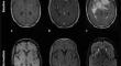
Results: We evaluated the baseline, intermediate, end-of-treatment and follow-up MRI data of 31 patients (9 living with HIV, 16 with solid organ transplantation and 6 with an autoimmune disease under chronic immunosuppressive therapy). At baseline, 23/30 (77%) patients had necrotic lesions with ring enhancement and 28% of the lesions were hemorrhagic. At the end of the first-line treatment, 12/28 (43%) patients could not be classified according to the IPCG criteria. Thirteen of 28 (46%) patients still harbored contrast enhancement, and 11/28 (39%) patients had persistent large necrotic lesions with a median diameter of 15 mm. These aspects were not associated with a pejorative outcome and progressively diminished during follow-up. Six patients relapsed; however, we failed to identify any neuroimaging risk factors on the end-of-treatment MRI.
Conclusion: In immunocompromised patients, PCNSLs often harbor alarming features on end-of-treatment MRI, with persistent contrast-enhanced lesions frequently observed. However, these aspects seemed to be related to the necrotic and hemorrhagic nature of the lesions and were not predictive of a pejorative outcome. Specific response criteria for this population are thereby proposed.
期刊介绍:
The Journal of Neuro-Oncology is a multi-disciplinary journal encompassing basic, applied, and clinical investigations in all research areas as they relate to cancer and the central nervous system. It provides a single forum for communication among neurologists, neurosurgeons, radiotherapists, medical oncologists, neuropathologists, neurodiagnosticians, and laboratory-based oncologists conducting relevant research. The Journal of Neuro-Oncology does not seek to isolate the field, but rather to focus the efforts of many disciplines in one publication through a format which pulls together these diverse interests. More than any other field of oncology, cancer of the central nervous system requires multi-disciplinary approaches. To alleviate having to scan dozens of journals of cell biology, pathology, laboratory and clinical endeavours, JNO is a periodical in which current, high-quality, relevant research in all aspects of neuro-oncology may be found.

 求助内容:
求助内容: 应助结果提醒方式:


