Fan Yang, Qiming He, Yanxia Wang, Siqi Zeng, Yingming Xu, Jing Ye, Yonghong He, Tian Guan, Zhe Wang, Jing Li
{"title":"Unsupervised stain augmentation enhanced glomerular instance segmentation on pathology images.","authors":"Fan Yang, Qiming He, Yanxia Wang, Siqi Zeng, Yingming Xu, Jing Ye, Yonghong He, Tian Guan, Zhe Wang, Jing Li","doi":"10.1007/s11548-024-03154-7","DOIUrl":null,"url":null,"abstract":"<p><strong>Purpose: </strong>In pathology images, different stains highlight different glomerular structures, so a supervised deep learning-based glomerular instance segmentation model trained on individual stains performs poorly on other stains. However, it is difficult to obtain a training set with multiple stains because the labeling of pathology images is very time-consuming and tedious. Therefore, in this paper, we proposed an unsupervised stain augmentation-based method for segmentation of glomerular instances.</p><p><strong>Methods: </strong>In this study, we successfully realized the conversion between different staining methods such as PAS, MT and PASM by contrastive unpaired translation (CUT), thus improving the staining diversity of the training set. Moreover, we replaced the backbone of mask R-CNN with swin transformer to further improve the efficiency of feature extraction and thus achieve better performance in instance segmentation task.</p><p><strong>Results: </strong>To validate the method presented in this paper, we constructed a dataset from 216 WSIs of the three stains in this study. After conducting in-depth experiments, we verified that the instance segmentation method based on stain augmentation outperforms existing methods across all metrics for PAS, PASM, and MT stains. Furthermore, ablation experiments are performed in this paper to further demonstrate the effectiveness of the proposed module.</p><p><strong>Conclusion: </strong>This study successfully demonstrated the potential of unsupervised stain augmentation to improve glomerular segmentation in pathology analysis. Future research could extend this approach to other complex segmentation tasks in the pathology image domain to further explore the potential of applying stain augmentation techniques in different domains of pathology image analysis.</p>","PeriodicalId":51251,"journal":{"name":"International Journal of Computer Assisted Radiology and Surgery","volume":" ","pages":"225-236"},"PeriodicalIF":2.3000,"publicationDate":"2025-02-01","publicationTypes":"Journal Article","fieldsOfStudy":null,"isOpenAccess":false,"openAccessPdf":"","citationCount":"0","resultStr":null,"platform":"Semanticscholar","paperid":null,"PeriodicalName":"International Journal of Computer Assisted Radiology and Surgery","FirstCategoryId":"5","ListUrlMain":"https://doi.org/10.1007/s11548-024-03154-7","RegionNum":3,"RegionCategory":"医学","ArticlePicture":[],"TitleCN":null,"AbstractTextCN":null,"PMCID":null,"EPubDate":"2024/6/7 0:00:00","PubModel":"Epub","JCR":"Q3","JCRName":"ENGINEERING, BIOMEDICAL","Score":null,"Total":0}
引用次数: 0
Abstract
Purpose: In pathology images, different stains highlight different glomerular structures, so a supervised deep learning-based glomerular instance segmentation model trained on individual stains performs poorly on other stains. However, it is difficult to obtain a training set with multiple stains because the labeling of pathology images is very time-consuming and tedious. Therefore, in this paper, we proposed an unsupervised stain augmentation-based method for segmentation of glomerular instances.
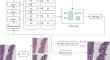
Methods: In this study, we successfully realized the conversion between different staining methods such as PAS, MT and PASM by contrastive unpaired translation (CUT), thus improving the staining diversity of the training set. Moreover, we replaced the backbone of mask R-CNN with swin transformer to further improve the efficiency of feature extraction and thus achieve better performance in instance segmentation task.
Results: To validate the method presented in this paper, we constructed a dataset from 216 WSIs of the three stains in this study. After conducting in-depth experiments, we verified that the instance segmentation method based on stain augmentation outperforms existing methods across all metrics for PAS, PASM, and MT stains. Furthermore, ablation experiments are performed in this paper to further demonstrate the effectiveness of the proposed module.
Conclusion: This study successfully demonstrated the potential of unsupervised stain augmentation to improve glomerular segmentation in pathology analysis. Future research could extend this approach to other complex segmentation tasks in the pathology image domain to further explore the potential of applying stain augmentation techniques in different domains of pathology image analysis.
期刊介绍:
The International Journal for Computer Assisted Radiology and Surgery (IJCARS) is a peer-reviewed journal that provides a platform for closing the gap between medical and technical disciplines, and encourages interdisciplinary research and development activities in an international environment.

 求助内容:
求助内容: 应助结果提醒方式:


