Faraz H Khan, Debbie Zhao, Jong-Won Ha, Sherif F Nagueh, Jens-Uwe Voigt, Allan L Klein, Einar Gude, Kaspar Broch, Nicholas Chan, Gina M Quill, Robert N Doughty, Alistair Young, Ji-Won Seo, Eusebio García-Izquierdo, Vanessa Moñivas-Palomero, Susana Mingo-Santos, Tom Kai Ming Wang, Stephanie Bezy, Nobuyuki Ohte, Helge Skulstad, Carmen C Beladan, Bogdan A Popescu, Shohei Kikuchi, Vasileios Panis, Erwan Donal, Espen W Remme, Martyn P Nash, Otto A Smiseth
{"title":"Evaluation of left ventricular filling pressure by echocardiography in patients with atrial fibrillation.","authors":"Faraz H Khan, Debbie Zhao, Jong-Won Ha, Sherif F Nagueh, Jens-Uwe Voigt, Allan L Klein, Einar Gude, Kaspar Broch, Nicholas Chan, Gina M Quill, Robert N Doughty, Alistair Young, Ji-Won Seo, Eusebio García-Izquierdo, Vanessa Moñivas-Palomero, Susana Mingo-Santos, Tom Kai Ming Wang, Stephanie Bezy, Nobuyuki Ohte, Helge Skulstad, Carmen C Beladan, Bogdan A Popescu, Shohei Kikuchi, Vasileios Panis, Erwan Donal, Espen W Remme, Martyn P Nash, Otto A Smiseth","doi":"10.1186/s44156-024-00048-x","DOIUrl":null,"url":null,"abstract":"<p><strong>Background: </strong>Echocardiography is widely used to evaluate left ventricular (LV) diastolic function in patients suspected of heart failure. For patients in sinus rhythm, a combination of several echocardiographic parameters can differentiate between normal and elevated LV filling pressure with good accuracy. However, there is no established echocardiographic approach for the evaluation of LV filling pressure in patients with atrial fibrillation. The objective of the present study was to determine if a combination of several echocardiographic and clinical parameters may be used to evaluate LV filling pressure in patients with atrial fibrillation.</p><p><strong>Results: </strong>In a multicentre study of 148 atrial fibrillation patients, several echocardiographic parameters were tested against invasively measured LV filling pressure as the reference method. No single parameter had sufficiently strong association with LV filling pressure to be recommended for clinical use. Based on univariate regression analysis in the present study, and evidence from existing literature, we developed a two-step algorithm for differentiation between normal and elevated LV filling pressure, defining values ≥ 15 mmHg as elevated. The parameters in the first step included the ratio between mitral early flow velocity and septal mitral annular velocity (septal E/e'), mitral E velocity, deceleration time of E, and peak tricuspid regurgitation velocity. Patients who could not be classified in the first step were tested in a second step by applying supplementary parameters, which included left atrial reservoir strain, pulmonary venous systolic/diastolic velocity ratio, and body mass index. This two-step algorithm classified patients as having either normal or elevated LV filling pressure with 75% accuracy and with 85% feasibility. Accuracy in EF ≥ 50% and EF < 50% was similar (75% and 76%).</p><p><strong>Conclusions: </strong>In patients with atrial fibrillation, no single echocardiographic parameter was sufficiently reliable to be used clinically to identify elevated LV filling pressure. An algorithm that combined several echocardiographic parameters and body mass index, however, was able to classify patients as having normal or elevated LV filling pressure with moderate accuracy and high feasibility.</p>","PeriodicalId":45749,"journal":{"name":"Echo Research and Practice","volume":"11 1","pages":"14"},"PeriodicalIF":2.4000,"publicationDate":"2024-06-03","publicationTypes":"Journal Article","fieldsOfStudy":null,"isOpenAccess":false,"openAccessPdf":"https://www.ncbi.nlm.nih.gov/pmc/articles/PMC11145766/pdf/","citationCount":"0","resultStr":null,"platform":"Semanticscholar","paperid":null,"PeriodicalName":"Echo Research and Practice","FirstCategoryId":"1085","ListUrlMain":"https://doi.org/10.1186/s44156-024-00048-x","RegionNum":0,"RegionCategory":null,"ArticlePicture":[],"TitleCN":null,"AbstractTextCN":null,"PMCID":null,"EPubDate":"","PubModel":"","JCR":"Q2","JCRName":"CARDIAC & CARDIOVASCULAR SYSTEMS","Score":null,"Total":0}
引用次数: 0
Abstract
Background: Echocardiography is widely used to evaluate left ventricular (LV) diastolic function in patients suspected of heart failure. For patients in sinus rhythm, a combination of several echocardiographic parameters can differentiate between normal and elevated LV filling pressure with good accuracy. However, there is no established echocardiographic approach for the evaluation of LV filling pressure in patients with atrial fibrillation. The objective of the present study was to determine if a combination of several echocardiographic and clinical parameters may be used to evaluate LV filling pressure in patients with atrial fibrillation.
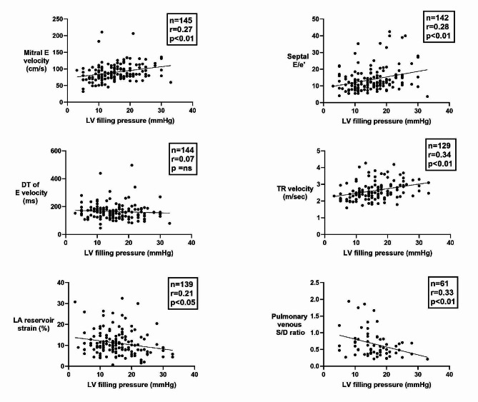
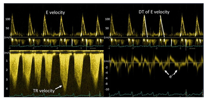
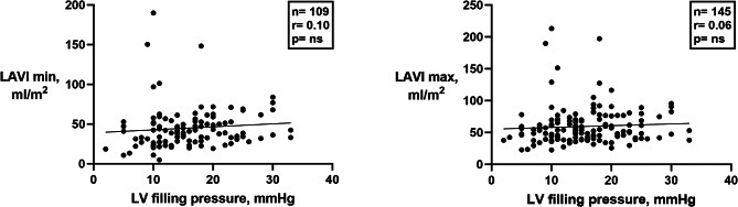
Results: In a multicentre study of 148 atrial fibrillation patients, several echocardiographic parameters were tested against invasively measured LV filling pressure as the reference method. No single parameter had sufficiently strong association with LV filling pressure to be recommended for clinical use. Based on univariate regression analysis in the present study, and evidence from existing literature, we developed a two-step algorithm for differentiation between normal and elevated LV filling pressure, defining values ≥ 15 mmHg as elevated. The parameters in the first step included the ratio between mitral early flow velocity and septal mitral annular velocity (septal E/e'), mitral E velocity, deceleration time of E, and peak tricuspid regurgitation velocity. Patients who could not be classified in the first step were tested in a second step by applying supplementary parameters, which included left atrial reservoir strain, pulmonary venous systolic/diastolic velocity ratio, and body mass index. This two-step algorithm classified patients as having either normal or elevated LV filling pressure with 75% accuracy and with 85% feasibility. Accuracy in EF ≥ 50% and EF < 50% was similar (75% and 76%).
Conclusions: In patients with atrial fibrillation, no single echocardiographic parameter was sufficiently reliable to be used clinically to identify elevated LV filling pressure. An algorithm that combined several echocardiographic parameters and body mass index, however, was able to classify patients as having normal or elevated LV filling pressure with moderate accuracy and high feasibility.
期刊介绍:
Echo Research and Practice aims to be the premier international journal for physicians, sonographers, nurses and other allied health professionals practising echocardiography and other cardiac imaging modalities. This open-access journal publishes quality clinical and basic research, reviews, videos, education materials and selected high-interest case reports and videos across all echocardiography modalities and disciplines, including paediatrics, anaesthetics, general practice, acute medicine and intensive care. Multi-modality studies primarily featuring the use of cardiac ultrasound in clinical practice, in association with Cardiac Computed Tomography, Cardiovascular Magnetic Resonance or Nuclear Cardiology are of interest. Topics include, but are not limited to: 2D echocardiography 3D echocardiography Comparative imaging techniques – CCT, CMR and Nuclear Cardiology Congenital heart disease, including foetal echocardiography Contrast echocardiography Critical care echocardiography Deformation imaging Doppler echocardiography Interventional echocardiography Intracardiac echocardiography Intraoperative echocardiography Prosthetic valves Stress echocardiography Technical innovations Transoesophageal echocardiography Valve disease.

 求助内容:
求助内容: 应助结果提醒方式:


