Reduced REST Expression in Neural Progenitor Cells, Adult Cortex, and Impaired REST Nuclear Translocation in the Prefrontal Cortex of Ts1Cje Mouse Model of Down Syndrome
{"title":"Reduced REST Expression in Neural Progenitor Cells, Adult Cortex, and Impaired REST Nuclear Translocation in the Prefrontal Cortex of Ts1Cje Mouse Model of Down Syndrome","authors":"Chong-Teik Lim, Xin-Jieh Lam, Arthini-Arrumugam Crystal, Tan Huang, Norhazlin Jusoh, Pike-See Cheah, King-Hwa Ling","doi":"10.1134/s1819712424010148","DOIUrl":null,"url":null,"abstract":"<h3 data-test=\"abstract-sub-heading\">Abstract</h3><p>Down syndrome (DS) is a common genetic disorder caused by trisomy of human chromosome 21. DS individuals have neurodevelopmental defects that lead to the manifestation of neurological and neuropsychiatric disorders. Repressor element-1 silencing transcription factor (REST) is the key epigenetic neuronal gene expression regulator. A comprehensive spatiotemporal profiling of <i>REST</i> expression is needed to understand its role in DS brain development. Therefore, we characterised REST targets in this study and profiled its expression using the brain samples from Ts1Cje, a mouse model exhibiting DS neuropathology. Over-representation analysis of Ts1Cje differentially expressed genes (DEGs) with mouse REST targets was performed. The cerebral cortex, hippocampus, and cerebellum of Ts1Cje and wildtype (WT) mice were procured at postnatal—P1, P15, P30, and P84 and embryonic—E14 and P1.5 development timepoints. RNAs from the brain tissues and cultured neurospheres were analysed with qPCR to determine the spatiotemporal profile of <i>Rest</i> expression. Western blot and immunohistochemistry (IHC) staining were performed to assess the level of REST expression and nuclear localisation. Over-representation analysis showed the Ts1Cje DEGs were significantly overlapped with mouse REST target genes. QPCR and Western blot analysis revealed a significant downregulation of <i>Rest</i> transcript in neurospheres and protein in Ts1Cje compared to WT. IHC staining showed REST perinuclear marginalisation and significantly reduced nuclear REST expression in the Ts1Cje prefrontal cortex. Loss of functional REST repression may lead to de-repression of DEGs in the Ts1Cje brain, potentially leading to various neuropathology seen in the Ts1Cje or DS brain.</p>","PeriodicalId":19119,"journal":{"name":"Neurochemical Journal","volume":"53 1","pages":""},"PeriodicalIF":0.5000,"publicationDate":"2024-05-24","publicationTypes":"Journal Article","fieldsOfStudy":null,"isOpenAccess":false,"openAccessPdf":"","citationCount":"0","resultStr":null,"platform":"Semanticscholar","paperid":null,"PeriodicalName":"Neurochemical Journal","FirstCategoryId":"3","ListUrlMain":"https://doi.org/10.1134/s1819712424010148","RegionNum":4,"RegionCategory":"医学","ArticlePicture":[],"TitleCN":null,"AbstractTextCN":null,"PMCID":null,"EPubDate":"","PubModel":"","JCR":"Q4","JCRName":"NEUROSCIENCES","Score":null,"Total":0}
引用次数: 0
Abstract
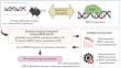
Down syndrome (DS) is a common genetic disorder caused by trisomy of human chromosome 21. DS individuals have neurodevelopmental defects that lead to the manifestation of neurological and neuropsychiatric disorders. Repressor element-1 silencing transcription factor (REST) is the key epigenetic neuronal gene expression regulator. A comprehensive spatiotemporal profiling of REST expression is needed to understand its role in DS brain development. Therefore, we characterised REST targets in this study and profiled its expression using the brain samples from Ts1Cje, a mouse model exhibiting DS neuropathology. Over-representation analysis of Ts1Cje differentially expressed genes (DEGs) with mouse REST targets was performed. The cerebral cortex, hippocampus, and cerebellum of Ts1Cje and wildtype (WT) mice were procured at postnatal—P1, P15, P30, and P84 and embryonic—E14 and P1.5 development timepoints. RNAs from the brain tissues and cultured neurospheres were analysed with qPCR to determine the spatiotemporal profile of Rest expression. Western blot and immunohistochemistry (IHC) staining were performed to assess the level of REST expression and nuclear localisation. Over-representation analysis showed the Ts1Cje DEGs were significantly overlapped with mouse REST target genes. QPCR and Western blot analysis revealed a significant downregulation of Rest transcript in neurospheres and protein in Ts1Cje compared to WT. IHC staining showed REST perinuclear marginalisation and significantly reduced nuclear REST expression in the Ts1Cje prefrontal cortex. Loss of functional REST repression may lead to de-repression of DEGs in the Ts1Cje brain, potentially leading to various neuropathology seen in the Ts1Cje or DS brain.
期刊介绍:
Neurochemical Journal (Neirokhimiya) provides a source for the communication of the latest findings in all areas of contemporary neurochemistry and other fields of relevance (including molecular biology, biochemistry, physiology, neuroimmunology, pharmacology) in an afford to expand our understanding of the functions of the nervous system. The journal presents papers on functional neurochemistry, nervous system receptors, neurotransmitters, myelin, chromaffin granules and other components of the nervous system, as well as neurophysiological and clinical aspects, behavioral reactions, etc. Relevant topics include structure and function of the nervous system proteins, neuropeptides, nucleic acids, nucleotides, lipids, and other biologically active components.
The journal is devoted to the rapid publication of regular papers containing the results of original research, reviews highlighting major developments in neurochemistry, short communications, new experimental studies that use neurochemical methodology, descriptions of new methods of value for neurochemistry, theoretical material suggesting novel principles and approaches to neurochemical problems, presentations of new hypotheses and significant findings, discussions, chronicles of congresses, meetings, and conferences with short presentations of the most sensational and timely reports, information on the activity of the Russian and International Neurochemical Societies, as well as advertisements of reagents and equipment.
 求助内容:
求助内容: 应助结果提醒方式:


