Automated multivolume placental reconstruction using three-dimensional power Doppler ultrasound and infrared camera tracking.

IF 6.1 1区 医学 Q1 ACOUSTICS
Ultrasound in Obstetrics & Gynecology Pub Date : 2025-05-01 Epub Date: 2025-04-03 DOI:10.1002/uog.27708
A Xue, R Hanly, D Luichareonkit, S Thomas, T Barber, A W Welsh
{"title":"Automated multivolume placental reconstruction using three-dimensional power Doppler ultrasound and infrared camera tracking.","authors":"A Xue, R Hanly, D Luichareonkit, S Thomas, T Barber, A W Welsh","doi":"10.1002/uog.27708","DOIUrl":null,"url":null,"abstract":"<p><strong>Objectives: </strong>Placental insufficiency contributes to many obstetric pathologies, but there is no bedside clinical tool to evaluate placental perfusion. We have developed a method to acquire multiple three-dimensional (3D) power Doppler (PD) ultrasound (US) volumes of placental vasculature, with infrared camera tracking of the precise spatial location of the transducer providing global coordinates. These volumes are reconstructed automatically ('stitched') into a model of the entire placenta. The purpose of this study was to evaluate the accuracy of automated reconstruction in an US phantom and to assess the feasibility of this technique in second-to-third-trimester human placentae.</p><p><strong>Methods: </strong>A custom-designed acrylic phantom was constructed with dimensions mimicking a third-trimester placenta, containing 12 rectangular cuboid towers of various heights submersed in tissue-mimicking solution. Multiple overlapping 3D-US volumes of this phantom were acquired using three different insonation angles and infrared camera tracking. Data were transformed into a 3D cartesian volume and stitched automatically into six 3D-US volumes, each covering the entire phantom, for each of the three different insonation angles. Reconstruction accuracy was evaluated by calculating local distance error (assessment of towers in overlapping US volumes to determine accuracy of stitching) and global distance error (subtraction of true measurements in phantom model from corresponding measurements in stitched 3D-US volumes). A single-center, cross-sectional feasibility study was then conducted in women with an uncomplicated second-to-third-trimester singleton pregnancy, with data obtained using standardized ultrasound settings. Multiple 3D PD-US and grayscale volumes of the placentae were acquired with infrared camera-tracked coordinates. Volumes were stitched to create a model of placental vasculature, and these were assessed for quality and repeatability of volume measurement.</p><p><strong>Results: </strong>Six entire phantom datasets were reconstructed at each of three insonation angles, giving a total of 18 extended phanom datasets. A median of nine 3D-US volumes required to reconstruct the entire phantom. Twelve towers per volume were assessed on three separate occasions, generating 648 datapoints. Of these datapoints, 67.1% were perfectly aligned. The mean local distance error was 2.92 (range, 0-25.51) mm. Measurements between towers of 120 distances in each stitched 3D-US volume (2160 distances in total) differed by an average of 1.51 (range, -4.78 to 4.23) mm from the true measurements in the phantom model. In the feasibility study, 17 participants were scanned, and 49 3D-US volume datasets acquired, with 92% reconstruction success per placental volume set and at least one complete volume being obtained per participant (100% participant achievability). The median volume acquisition and reconstruction time was 10 min. Reconstructed placental vasculature was assessed qualitatively to be present, continuous and detailed throughout. Volume measurement of entire segmented placentae was highly repeatable (intraclass correlation coefficient, 0.96 (95% CI, 0.89-0.99)).</p><p><strong>Conclusion: </strong>We present an automated method to model the entire structure and vasculature of second-to-third-trimester placentae using multiple 3D PD-US volumes, with verified accuracy and clinical feasibility. This study builds a foundation on which to develop a practical screening tool for detecting placental insufficiency and has the potential for expansion to evaluation of adult organ perfusion. © 2024 The Author(s). Ultrasound in Obstetrics & Gynecology published by John Wiley & Sons Ltd on behalf of International Society of Ultrasound in Obstetrics and Gynecology.</p>","PeriodicalId":23454,"journal":{"name":"Ultrasound in Obstetrics & Gynecology","volume":" ","pages":"624-632"},"PeriodicalIF":6.1000,"publicationDate":"2025-05-01","publicationTypes":"Journal Article","fieldsOfStudy":null,"isOpenAccess":false,"openAccessPdf":"https://www.ncbi.nlm.nih.gov/pmc/articles/PMC12047681/pdf/","citationCount":"0","resultStr":null,"platform":"Semanticscholar","paperid":null,"PeriodicalName":"Ultrasound in Obstetrics & Gynecology","FirstCategoryId":"3","ListUrlMain":"https://doi.org/10.1002/uog.27708","RegionNum":1,"RegionCategory":"医学","ArticlePicture":[],"TitleCN":null,"AbstractTextCN":null,"PMCID":null,"EPubDate":"2025/4/3 0:00:00","PubModel":"Epub","JCR":"Q1","JCRName":"ACOUSTICS","Score":null,"Total":0}
引用次数: 0

Abstract

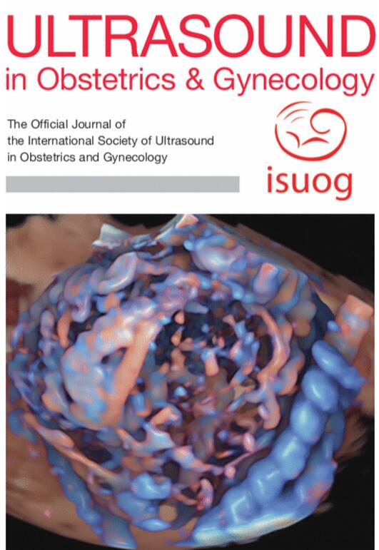
Objectives: Placental insufficiency contributes to many obstetric pathologies, but there is no bedside clinical tool to evaluate placental perfusion. We have developed a method to acquire multiple three-dimensional (3D) power Doppler (PD) ultrasound (US) volumes of placental vasculature, with infrared camera tracking of the precise spatial location of the transducer providing global coordinates. These volumes are reconstructed automatically ('stitched') into a model of the entire placenta. The purpose of this study was to evaluate the accuracy of automated reconstruction in an US phantom and to assess the feasibility of this technique in second-to-third-trimester human placentae.

Methods: A custom-designed acrylic phantom was constructed with dimensions mimicking a third-trimester placenta, containing 12 rectangular cuboid towers of various heights submersed in tissue-mimicking solution. Multiple overlapping 3D-US volumes of this phantom were acquired using three different insonation angles and infrared camera tracking. Data were transformed into a 3D cartesian volume and stitched automatically into six 3D-US volumes, each covering the entire phantom, for each of the three different insonation angles. Reconstruction accuracy was evaluated by calculating local distance error (assessment of towers in overlapping US volumes to determine accuracy of stitching) and global distance error (subtraction of true measurements in phantom model from corresponding measurements in stitched 3D-US volumes). A single-center, cross-sectional feasibility study was then conducted in women with an uncomplicated second-to-third-trimester singleton pregnancy, with data obtained using standardized ultrasound settings. Multiple 3D PD-US and grayscale volumes of the placentae were acquired with infrared camera-tracked coordinates. Volumes were stitched to create a model of placental vasculature, and these were assessed for quality and repeatability of volume measurement.

Results: Six entire phantom datasets were reconstructed at each of three insonation angles, giving a total of 18 extended phanom datasets. A median of nine 3D-US volumes required to reconstruct the entire phantom. Twelve towers per volume were assessed on three separate occasions, generating 648 datapoints. Of these datapoints, 67.1% were perfectly aligned. The mean local distance error was 2.92 (range, 0-25.51) mm. Measurements between towers of 120 distances in each stitched 3D-US volume (2160 distances in total) differed by an average of 1.51 (range, -4.78 to 4.23) mm from the true measurements in the phantom model. In the feasibility study, 17 participants were scanned, and 49 3D-US volume datasets acquired, with 92% reconstruction success per placental volume set and at least one complete volume being obtained per participant (100% participant achievability). The median volume acquisition and reconstruction time was 10 min. Reconstructed placental vasculature was assessed qualitatively to be present, continuous and detailed throughout. Volume measurement of entire segmented placentae was highly repeatable (intraclass correlation coefficient, 0.96 (95% CI, 0.89-0.99)).

Conclusion: We present an automated method to model the entire structure and vasculature of second-to-third-trimester placentae using multiple 3D PD-US volumes, with verified accuracy and clinical feasibility. This study builds a foundation on which to develop a practical screening tool for detecting placental insufficiency and has the potential for expansion to evaluation of adult organ perfusion. © 2024 The Author(s). Ultrasound in Obstetrics & Gynecology published by John Wiley & Sons Ltd on behalf of International Society of Ultrasound in Obstetrics and Gynecology.

利用三维动力多普勒超声和红外摄像跟踪技术自动重建多容积胎盘。
目的:胎盘功能不全可导致多种产科病症,但目前尚无床旁临床工具来评估胎盘灌注情况。我们开发了一种方法,通过红外相机跟踪提供全局坐标,获取胎盘血管的多个三维动力多普勒超声(3D PD-US)体积。这些数据会自动重建("拼接")成整个胎盘的模型。这项研究的目的是评估美国模型中自动重建的准确性,并将此技术应用于人类胎盘:方法:按照模仿妊娠三个月胎盘的尺寸定制了一个丙烯酸模型,其中包含 12 个高度不等的四边形塔,并浸没在组织模拟溶液中。利用红外相机跟踪技术,从三个不同的采集角度采集了该模型的三维超声体积。数据被转换成三维直角坐标体并自动拼接。采用标准化产科设置,对无并发症的第二至第三孕期单胎妊娠进行了单中心横断面可行性研究。胎盘的多个三维 PD-US 和灰度体积是通过红外相机跟踪坐标获得的。对胎盘体积进行拼接,以创建胎盘血管模型:在 3 个容积角度下分别重建了 6 个模型数据集,所需的容积中位数为 9 个。在 648 个数据点中,有 66.7% 实现了完美的容积对齐。容积错位的平均距离误差为 2.92 毫米。每个拼接卷中 210 个距离的测量值(总计 2160 个距离)与真实测量值平均相差 1.51 毫米。这些结果与最近的文献相比毫不逊色,尽管使用的是更大的模型。对 17 名参与者进行了扫描,每组胎盘容积的重建成功率为 92%,参与者的成功率为 100%。中位重建时间为 10 分钟。经定性评估,胎盘血管在整个扫描过程中都是存在的、连续的和详细的。整个分割胎盘的体积测量具有很高的重复性(ICC 0.96):我们提出了一种自动方法来模拟第二至第三孕期胎盘的整体结构和脉管系统,其准确性和临床可行性已通过灰度和功率多普勒的验证。这项研究为开发实用的胎盘功能不全筛查工具奠定了基础,并可扩展到成人器官灌注评估。本文受版权保护。保留所有权利。
本文章由计算机程序翻译,如有差异,请以英文原文为准。
求助全文
约1分钟内获得全文 求助全文
来源期刊
CiteScore
12.30
自引率
14.10%
发文量
891
审稿时长
1 months
期刊介绍: Ultrasound in Obstetrics & Gynecology (UOG) is the official journal of the International Society of Ultrasound in Obstetrics and Gynecology (ISUOG) and is considered the foremost international peer-reviewed journal in the field. It publishes cutting-edge research that is highly relevant to clinical practice, which includes guidelines, expert commentaries, consensus statements, original articles, and systematic reviews. UOG is widely recognized and included in prominent abstract and indexing databases such as Index Medicus and Current Contents.
×
引用
GB/T 7714-2015
复制
MLA
复制
APA
复制
导出至
BibTeX EndNote RefMan NoteFirst NoteExpress
×
提示
您的信息不完整,为了账户安全,请先补充。
现在去补充
×
提示
您因"违规操作"
具体请查看互助需知
我知道了
×
提示
确定
请完成安全验证×
copy
已复制链接
快去分享给好友吧!
我知道了
右上角分享
点击右上角分享
0
联系我们:info@booksci.cn Book学术提供免费学术资源搜索服务,方便国内外学者检索中英文文献。致力于提供最便捷和优质的服务体验。 Copyright © 2023 布克学术 All rights reserved.
京ICP备2023020795号-1
ghs 京公网安备 11010802042870号
Book学术文献互助
Book学术文献互助群
群 号:481959085
Book学术官方微信