Radiomics analysis of ultrasound images to discriminate between benign and malignant adnexal masses with solid morphology on ultrasound.

IF 6.1 1区 医学 Q1 ACOUSTICS
Ultrasound in Obstetrics & Gynecology Pub Date : 2025-03-01 Epub Date: 2025-02-02 DOI:10.1002/uog.27680
F Moro, M Vagni, H E Tran, F Bernardini, F Mascilini, F Ciccarone, C Nero, D Giannarelli, L Boldrini, A Fagotti, G Scambia, L Valentin, A C Testa
{"title":"Radiomics analysis of ultrasound images to discriminate between benign and malignant adnexal masses with solid morphology on ultrasound.","authors":"F Moro, M Vagni, H E Tran, F Bernardini, F Mascilini, F Ciccarone, C Nero, D Giannarelli, L Boldrini, A Fagotti, G Scambia, L Valentin, A C Testa","doi":"10.1002/uog.27680","DOIUrl":null,"url":null,"abstract":"<p><strong>Objective: </strong>The primary aim was to identify radiomics ultrasound features that can distinguish between benign and malignant adnexal masses with solid ultrasound morphology, and between primary malignant (including borderline and primary invasive) and metastatic solid ovarian masses, and to develop ultrasound-based machine learning models that include radiomics features to discriminate between benign and malignant solid adnexal masses. The secondary aim was to compare the discrimination performance of our newly developed radiomics models with that of the Assessment of Different NEoplasias in the adneXa (ADNEX) model and that of subjective assessment by an experienced ultrasound examiner.</p><p><strong>Methods: </strong>This was a retrospective, observational single-center study conducted at Fondazione Policlinico Universitario A. Gemelli IRCC, in Rome, Italy. Included were patients with a histological diagnosis of an adnexal tumor with solid morphology according to International Ovarian Tumor Analysis (IOTA) terminology at preoperative ultrasound examination performed in 2014-2020, who were managed with surgery. The patient cohort was split randomly into training and validation sets at a ratio of 70:30 and with the same proportion of benign and malignant tumors in the two subsets, with malignant tumors including borderline, primary invasive and metastatic tumors. We extracted 68 radiomics features, belonging to two different families: intensity-based statistical features and textural features. Models to predict malignancy were built based on a random forest classifier, fine-tuned using 5-fold cross-validation over the training set, and tested on the held-out validation set. The variables used in model-building were patient age and radiomics features that were statistically significantly different between benign and malignant adnexal masses and assessed as not redundant based on the Pearson correlation coefficient. We evaluated the discriminative ability of the models and compared it to that of the ADNEX model and that of subjective assessment by an experienced ultrasound examiner using the area under the receiver-operating-characteristics curve (AUC) and classification performance by calculating sensitivity and specificity.</p><p><strong>Results: </strong>In total, 326 patients were included and 775 preoperative ultrasound images were analyzed. Of the 68 radiomics features extracted, 52 differed statistically significantly between benign and malignant tumors in the training set, and 18 uncorrelated features were selected for inclusion in model-building. The same 52 radiomics features differed significantly between benign, primary malignant and metastatic tumors. However, the values of the features manifested overlapped between primary malignant and metastatic tumors and did not differ significantly between them. In the validation set, 25/98 (25.5%) tumors were benign and 73/98 (74.5%) were malignant (6 borderline, 57 primary invasive, 10 metastatic). In the validation set, a model including only radiomics features had an AUC of 0.80, sensitivity of 0.78 and specificity of 0.76 at an optimal cut-off for risk of malignancy of 68%, based on Youden's index. The corresponding results for a model including age and radiomics features were AUC of 0.79, sensitivity of 0.86 and specificity of 0.56 (cut-off 60%, based on Youden's index), while those of the ADNEX model were AUC of 0.88, sensitivity of 0.99 and specificity of 0.64 (at a 20% risk-of-malignancy cut-off). Subjective assessment had a sensitivity of 0.99 and specificity of 0.72.</p><p><strong>Conclusions: </strong>Our radiomics model had moderate discriminative ability on internal validation and the addition of age to this model did not improve its performance. Even though our radiomics models had discriminative ability inferior to that of the ADNEX model, our results are sufficiently promising to justify continued development of radiomics analysis of ultrasound images of adnexal masses. © 2024 The Author(s). Ultrasound in Obstetrics & Gynecology published by John Wiley & Sons Ltd on behalf of International Society of Ultrasound in Obstetrics and Gynecology.</p>","PeriodicalId":23454,"journal":{"name":"Ultrasound in Obstetrics & Gynecology","volume":" ","pages":"353-363"},"PeriodicalIF":6.1000,"publicationDate":"2025-03-01","publicationTypes":"Journal Article","fieldsOfStudy":null,"isOpenAccess":false,"openAccessPdf":"https://www.ncbi.nlm.nih.gov/pmc/articles/PMC11872347/pdf/","citationCount":"0","resultStr":null,"platform":"Semanticscholar","paperid":null,"PeriodicalName":"Ultrasound in Obstetrics & Gynecology","FirstCategoryId":"3","ListUrlMain":"https://doi.org/10.1002/uog.27680","RegionNum":1,"RegionCategory":"医学","ArticlePicture":[],"TitleCN":null,"AbstractTextCN":null,"PMCID":null,"EPubDate":"2025/2/2 0:00:00","PubModel":"Epub","JCR":"Q1","JCRName":"ACOUSTICS","Score":null,"Total":0}
引用次数: 0

Abstract

Objective: The primary aim was to identify radiomics ultrasound features that can distinguish between benign and malignant adnexal masses with solid ultrasound morphology, and between primary malignant (including borderline and primary invasive) and metastatic solid ovarian masses, and to develop ultrasound-based machine learning models that include radiomics features to discriminate between benign and malignant solid adnexal masses. The secondary aim was to compare the discrimination performance of our newly developed radiomics models with that of the Assessment of Different NEoplasias in the adneXa (ADNEX) model and that of subjective assessment by an experienced ultrasound examiner.

Methods: This was a retrospective, observational single-center study conducted at Fondazione Policlinico Universitario A. Gemelli IRCC, in Rome, Italy. Included were patients with a histological diagnosis of an adnexal tumor with solid morphology according to International Ovarian Tumor Analysis (IOTA) terminology at preoperative ultrasound examination performed in 2014-2020, who were managed with surgery. The patient cohort was split randomly into training and validation sets at a ratio of 70:30 and with the same proportion of benign and malignant tumors in the two subsets, with malignant tumors including borderline, primary invasive and metastatic tumors. We extracted 68 radiomics features, belonging to two different families: intensity-based statistical features and textural features. Models to predict malignancy were built based on a random forest classifier, fine-tuned using 5-fold cross-validation over the training set, and tested on the held-out validation set. The variables used in model-building were patient age and radiomics features that were statistically significantly different between benign and malignant adnexal masses and assessed as not redundant based on the Pearson correlation coefficient. We evaluated the discriminative ability of the models and compared it to that of the ADNEX model and that of subjective assessment by an experienced ultrasound examiner using the area under the receiver-operating-characteristics curve (AUC) and classification performance by calculating sensitivity and specificity.

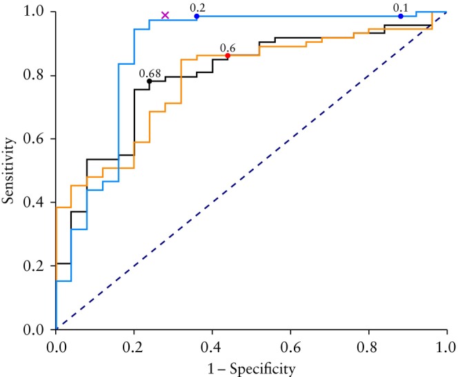
Results: In total, 326 patients were included and 775 preoperative ultrasound images were analyzed. Of the 68 radiomics features extracted, 52 differed statistically significantly between benign and malignant tumors in the training set, and 18 uncorrelated features were selected for inclusion in model-building. The same 52 radiomics features differed significantly between benign, primary malignant and metastatic tumors. However, the values of the features manifested overlapped between primary malignant and metastatic tumors and did not differ significantly between them. In the validation set, 25/98 (25.5%) tumors were benign and 73/98 (74.5%) were malignant (6 borderline, 57 primary invasive, 10 metastatic). In the validation set, a model including only radiomics features had an AUC of 0.80, sensitivity of 0.78 and specificity of 0.76 at an optimal cut-off for risk of malignancy of 68%, based on Youden's index. The corresponding results for a model including age and radiomics features were AUC of 0.79, sensitivity of 0.86 and specificity of 0.56 (cut-off 60%, based on Youden's index), while those of the ADNEX model were AUC of 0.88, sensitivity of 0.99 and specificity of 0.64 (at a 20% risk-of-malignancy cut-off). Subjective assessment had a sensitivity of 0.99 and specificity of 0.72.

Conclusions: Our radiomics model had moderate discriminative ability on internal validation and the addition of age to this model did not improve its performance. Even though our radiomics models had discriminative ability inferior to that of the ADNEX model, our results are sufficiently promising to justify continued development of radiomics analysis of ultrasound images of adnexal masses. © 2024 The Author(s). Ultrasound in Obstetrics & Gynecology published by John Wiley & Sons Ltd on behalf of International Society of Ultrasound in Obstetrics and Gynecology.

对超声图像进行放射组学分析,以区分具有实性超声形态的良性和恶性附件肿块。
目的我们的主要目的是确定可区分超声形态为实性的良性和恶性附件肿块以及原发性浸润性和转移性实性卵巢肿块的放射学超声特征,并开发包含放射学特征的超声机器学习模型,以区分良性和恶性实性附件肿块。我们的第二个目的是比较我们的放射组学模型与 ADNEX 模型的诊断性能,以及经验丰富的超声波检查员的主观评估:这是一项单中心回顾性观察研究。方法:这是一项回顾性观察性单中心研究,研究对象包括在 2014 年至 2021 年期间接受术前超声检查并经组织学诊断为实性形态的附件肿瘤患者。患者队列按 70:30 的比例分为训练集和验证集,两个子集中良性肿瘤和恶性肿瘤(边缘性、原发浸润性和转移性)的比例相同。提取的放射学特征分为两个不同的系列:基于强度的统计特征和纹理特征。预测恶性程度的模型是基于随机森林分类器建立的,在训练集上使用 5 倍交叉验证进行微调,并在保留的验证集上进行测试。建立模型时使用的变量包括患者的年龄,以及良性和恶性附件肿块之间存在显著统计学差异的放射学特征(Wilcoxon-Mann-Whitney 检验,并对多重比较进行本杰明-霍奇伯格校正),并根据皮尔逊相关系数评估这些特征是否多余。我们用接收者操作特征曲线下面积(AUC)来描述分辨能力,用灵敏度和特异性来描述分类效果:结果:共确定了 326 名患者,分析了 775 张术前超声图像。提取了 68 个放射学特征,其中 52 个特征在训练集中的良性肿瘤和恶性肿瘤之间存在显著的统计学差异,18 个特征被选中用于建立模型。这 52 个放射学特征在良性肿瘤、原发性浸润性恶性肿瘤和转移性肿瘤之间存在明显的统计学差异。不过,原发性恶性肿瘤和转移性肿瘤的特征值有重叠,在统计学上没有明显差异。在验证集中,25/98(25.5%)个肿瘤为良性,73/98(74.5%)个肿瘤为恶性(6 个边缘性肿瘤、57 个原发性浸润性肿瘤和 10 个转移性肿瘤)。在验证集中,仅包括放射组学特征的模型的AUC为0.80,在最佳恶性风险临界值(根据尤登指数为68%)时,灵敏度为78%,特异度为76%。包括年龄和放射组学特征在内的模型的相应结果分别为 0.79、86% 和 56%(根据尤登方法,临界值为 60%),而 ADNEX 模型的相应结果分别为 0.88、99% 和 64%(恶性风险临界值为 20%)。主观评估的灵敏度为 99%,特异性为 72%:尽管我们的放射组学模型的鉴别能力不如ADNEX模型,但我们的结果还是很有希望的,足以证明继续发展附件肿块超声图像的放射组学分析是有必要的。本文受版权保护。保留所有权利。
本文章由计算机程序翻译,如有差异,请以英文原文为准。
求助全文
约1分钟内获得全文 求助全文
来源期刊
CiteScore
12.30
自引率
14.10%
发文量
891
审稿时长
1 months
期刊介绍: Ultrasound in Obstetrics & Gynecology (UOG) is the official journal of the International Society of Ultrasound in Obstetrics and Gynecology (ISUOG) and is considered the foremost international peer-reviewed journal in the field. It publishes cutting-edge research that is highly relevant to clinical practice, which includes guidelines, expert commentaries, consensus statements, original articles, and systematic reviews. UOG is widely recognized and included in prominent abstract and indexing databases such as Index Medicus and Current Contents.
×
引用
GB/T 7714-2015
复制
MLA
复制
APA
复制
导出至
BibTeX EndNote RefMan NoteFirst NoteExpress
×
提示
您的信息不完整,为了账户安全,请先补充。
现在去补充
×
提示
您因"违规操作"
具体请查看互助需知
我知道了
×
提示
确定
请完成安全验证×
copy
已复制链接
快去分享给好友吧!
我知道了
右上角分享
点击右上角分享
0
联系我们:info@booksci.cn Book学术提供免费学术资源搜索服务,方便国内外学者检索中英文文献。致力于提供最便捷和优质的服务体验。 Copyright © 2023 布克学术 All rights reserved.
京ICP备2023020795号-1
ghs 京公网安备 11010802042870号
Book学术文献互助
Book学术文献互助群
群 号:481959085
Book学术官方微信