Jorge Cebrián, Victor Martínez, Pablo Hernández, Dora B Krimer, María-Luisa Martínez-Robles, Jorge B Schvartzman, María José Fernández-Nestosa
{"title":"Electrophoretic Mobility Assay to Separate Supercoiled, Catenated, and Knotted DNA Molecules.","authors":"Jorge Cebrián, Victor Martínez, Pablo Hernández, Dora B Krimer, María-Luisa Martínez-Robles, Jorge B Schvartzman, María José Fernández-Nestosa","doi":"10.21769/BioProtoc.4983","DOIUrl":null,"url":null,"abstract":"<p><p>Two-dimensional (2D) agarose gel electrophoresis is the method of choice to analyze DNA topology. The possibility to use <i>E. coli</i> strains with different genetic backgrounds in combination with nicking enzymes and different concentrations of norfloxacin improves the resolution of 2D gels to study the electrophoretic behavior of three different families of DNA topoisomers: supercoiled DNA molecules, post-replicative catenanes, and knotted DNA molecules. Here, we describe the materials and procedures required to optimize their separation by 2D gels. Understanding the differences in their electrophoretic behavior can help explain some important physical characteristics of these different types of DNA topoisomers. Key features • Preparative method to enrich DNA samples of supercoiled, catenated, and knotted families of topoisomers, later analyzed by 2D gels (or other techniques, e.g., microscopy). • 2D gels facilitate the separation of the topoisomers of any given circular DNA molecule. • Separation of DNA molecules with the same molecular masses but different shapes can be optimized by modifying the conditions of 2D gels. • Evaluating the roles of electric field and agarose concentration on the electrophoretic mobility of DNA topoisomers sheds light on their physical characteristics.</p>","PeriodicalId":93907,"journal":{"name":"Bio-protocol","volume":"14 9","pages":"e4983"},"PeriodicalIF":1.1000,"publicationDate":"2024-05-05","publicationTypes":"Journal Article","fieldsOfStudy":null,"isOpenAccess":false,"openAccessPdf":"https://www.ncbi.nlm.nih.gov/pmc/articles/PMC11082789/pdf/","citationCount":"0","resultStr":null,"platform":"Semanticscholar","paperid":null,"PeriodicalName":"Bio-protocol","FirstCategoryId":"1085","ListUrlMain":"https://doi.org/10.21769/BioProtoc.4983","RegionNum":0,"RegionCategory":null,"ArticlePicture":[],"TitleCN":null,"AbstractTextCN":null,"PMCID":null,"EPubDate":"","PubModel":"","JCR":"Q3","JCRName":"BIOLOGY","Score":null,"Total":0}
引用次数: 0
Abstract
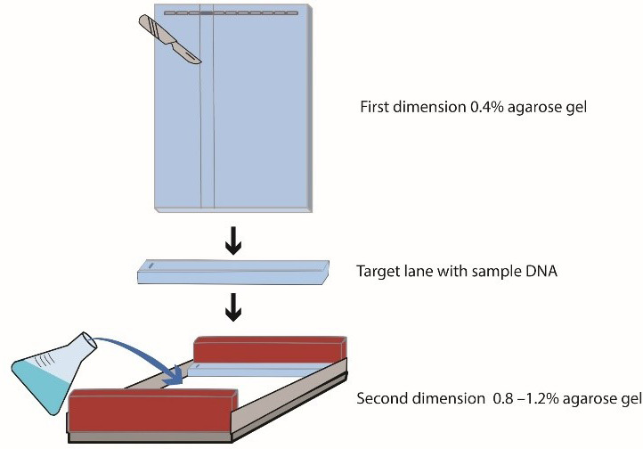
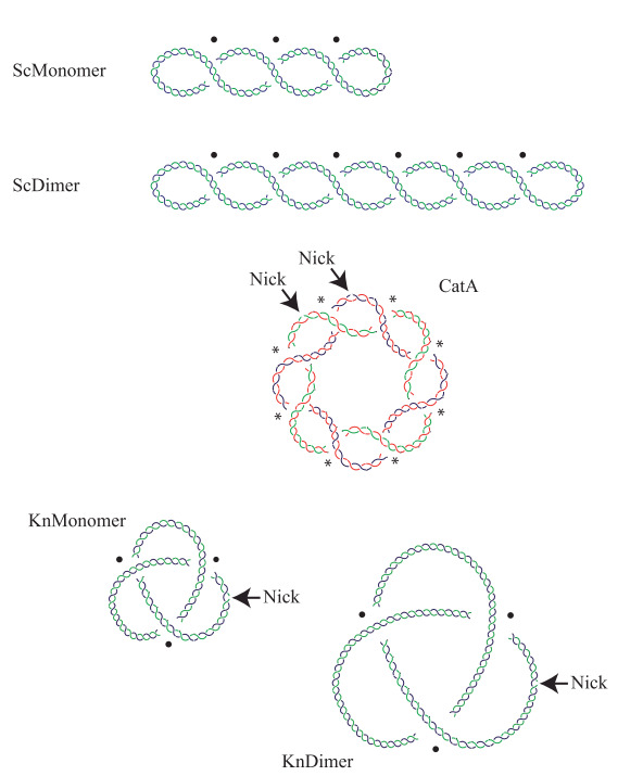
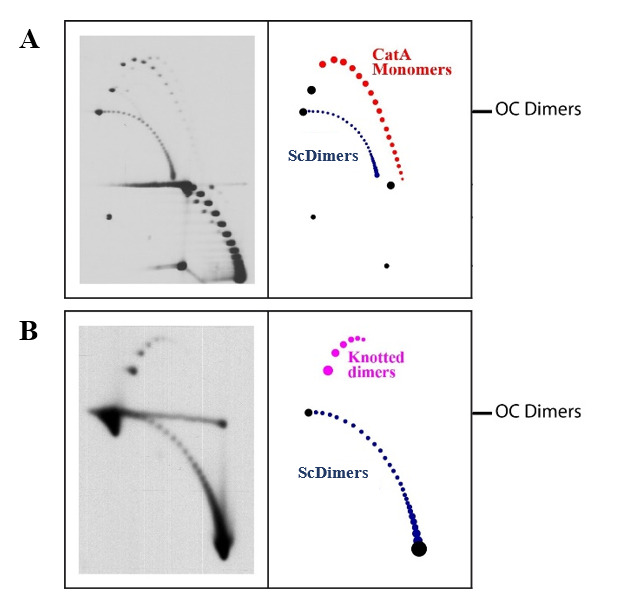
Two-dimensional (2D) agarose gel electrophoresis is the method of choice to analyze DNA topology. The possibility to use E. coli strains with different genetic backgrounds in combination with nicking enzymes and different concentrations of norfloxacin improves the resolution of 2D gels to study the electrophoretic behavior of three different families of DNA topoisomers: supercoiled DNA molecules, post-replicative catenanes, and knotted DNA molecules. Here, we describe the materials and procedures required to optimize their separation by 2D gels. Understanding the differences in their electrophoretic behavior can help explain some important physical characteristics of these different types of DNA topoisomers. Key features • Preparative method to enrich DNA samples of supercoiled, catenated, and knotted families of topoisomers, later analyzed by 2D gels (or other techniques, e.g., microscopy). • 2D gels facilitate the separation of the topoisomers of any given circular DNA molecule. • Separation of DNA molecules with the same molecular masses but different shapes can be optimized by modifying the conditions of 2D gels. • Evaluating the roles of electric field and agarose concentration on the electrophoretic mobility of DNA topoisomers sheds light on their physical characteristics.
二维(2D)琼脂糖凝胶电泳是分析 DNA 拓扑的首选方法。使用具有不同遗传背景的大肠杆菌菌株,结合切分酶和不同浓度的诺氟沙星,可以提高二维凝胶的分辨率,从而研究三种不同DNA拓扑异构体家族的电泳行为:超卷曲DNA分子、复制后双链DNA分子和打结DNA分子。在此,我们介绍了优化二维凝胶分离所需的材料和程序。了解它们电泳行为的差异有助于解释这些不同类型 DNA 拓扑异构体的一些重要物理特征。主要特点 - 用制备方法富集超卷曲、卡氏体和打结拓扑异构体系列的 DNA 样品,然后用二维凝胶(或其他技术,如显微镜)进行分析。- 二维凝胶有助于分离任何给定环状 DNA 分子的拓扑异构体。- 通过改变二维凝胶的条件,可以优化分子质量相同但形状不同的 DNA 分子的分离。- 评估电场和琼脂糖浓度对 DNA 拓扑异构体电泳迁移率的作用,有助于了解它们的物理特性。

 求助内容:
求助内容: 应助结果提醒方式:


