A. S. Goloveshkin, E. S. Kulikova, R. A. Novikov, A. V. Vologzhanina, A. A. Korlyukov
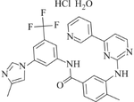
{"title":"Crystal Structure of Nilotinib Hydrochloride Monohydrate According to Powder X-Ray Diffraction Data","authors":"A. S. Goloveshkin, E. S. Kulikova, R. A. Novikov, A. V. Vologzhanina, A. A. Korlyukov","doi":"10.1134/S0022476624030132","DOIUrl":null,"url":null,"abstract":"<p>The crystal structure of nilotinib hydrochloride monohydrate, being an active pharmaceutical substance of the drug <i>Tasigna</i> for chronic myeloid leukemia, is determined by powder X-ray diffraction (XRD) using a synchrotron radiation source of the National Research Center “Kurchatov Institute”. The molecular conformation of the cation in the crystal differs from that in the initial nilotinib molecule and its solvates due to the rotation of substituents about single C–N, C–O, and C–C bonds. In the crystal, the nilotinib molecule is protonated at the nitrogen atom of the imidazolium heterocycle and involved in the N–H…N, N–H…O, and N–H…Cl hydrogen bonds with another cation, water molecule, and anion respectively. The resulting structure is determined with high accuracy. Errors in bond lengths are only slightly worse than those for the structures determined from single crystal XRD data. In addition, one of the best values of the half uncertainty window (HUW) is achieved in the refinement. The importance of this parameter is considered in the article. The structure agrees with the solid-state NMR data. The comparison of the results with the solution NMR spectroscopy data reveals noticeable changes in the nilotinib molecular structure during crystallization.</p>","PeriodicalId":668,"journal":{"name":"Journal of Structural Chemistry","volume":"65 3","pages":"585 - 595"},"PeriodicalIF":1.2000,"publicationDate":"2024-04-30","publicationTypes":"Journal Article","fieldsOfStudy":null,"isOpenAccess":false,"openAccessPdf":"","citationCount":"0","resultStr":null,"platform":"Semanticscholar","paperid":null,"PeriodicalName":"Journal of Structural Chemistry","FirstCategoryId":"92","ListUrlMain":"https://link.springer.com/article/10.1134/S0022476624030132","RegionNum":4,"RegionCategory":"化学","ArticlePicture":[],"TitleCN":null,"AbstractTextCN":null,"PMCID":null,"EPubDate":"","PubModel":"","JCR":"Q4","JCRName":"CHEMISTRY, INORGANIC & NUCLEAR","Score":null,"Total":0}
引用次数: 0
Abstract
The crystal structure of nilotinib hydrochloride monohydrate, being an active pharmaceutical substance of the drug Tasigna for chronic myeloid leukemia, is determined by powder X-ray diffraction (XRD) using a synchrotron radiation source of the National Research Center “Kurchatov Institute”. The molecular conformation of the cation in the crystal differs from that in the initial nilotinib molecule and its solvates due to the rotation of substituents about single C–N, C–O, and C–C bonds. In the crystal, the nilotinib molecule is protonated at the nitrogen atom of the imidazolium heterocycle and involved in the N–H…N, N–H…O, and N–H…Cl hydrogen bonds with another cation, water molecule, and anion respectively. The resulting structure is determined with high accuracy. Errors in bond lengths are only slightly worse than those for the structures determined from single crystal XRD data. In addition, one of the best values of the half uncertainty window (HUW) is achieved in the refinement. The importance of this parameter is considered in the article. The structure agrees with the solid-state NMR data. The comparison of the results with the solution NMR spectroscopy data reveals noticeable changes in the nilotinib molecular structure during crystallization.
期刊介绍:
Journal is an interdisciplinary publication covering all aspects of structural chemistry, including the theory of molecular structure and chemical bond; the use of physical methods to study the electronic and spatial structure of chemical species; structural features of liquids, solutions, surfaces, supramolecular systems, nano- and solid materials; and the crystal structure of solids.


 求助内容:
求助内容: 应助结果提醒方式:


