Nima Gilani, Artem Mikheev, Inge M Brinkmann, Malika Kumbella, James S Babb, Dibash Basukala, Andreas Wetscherek, Thomas Benkert, Hersh Chandarana, Eric E Sigmund
{"title":"Spatial profiling of in vivo diffusion-weighted MRI parameters in the healthy human kidney.","authors":"Nima Gilani, Artem Mikheev, Inge M Brinkmann, Malika Kumbella, James S Babb, Dibash Basukala, Andreas Wetscherek, Thomas Benkert, Hersh Chandarana, Eric E Sigmund","doi":"10.1007/s10334-024-01159-6","DOIUrl":null,"url":null,"abstract":"<p><strong>Objective: </strong>Diffusion-weighted MRI is a technique that can infer microstructural and microcirculatory features from biological tissue, with particular application to renal tissue. There is extensive literature on diffusion tensor imaging (DTI) of anisotropy in the renal medulla, intravoxel incoherent motion (IVIM) measurements separating microstructural from microcirculation effects, and combinations of the two. However, interpretation of these features and adaptation of more specific models remains an ongoing challenge. One input to this process is a whole organ distillation of corticomedullary contrast of diffusion metrics, as has been explored for other renal biomarkers.</p><p><strong>Materials and methods: </strong>In this work, we probe the spatial dependence of diffusion MRI metrics with concentrically layered segmentation in 11 healthy kidneys at 3 T. The metrics include those from DTI, IVIM, a combined approach titled \"REnal Flow and Microstructure AnisotroPy (REFMAP)\", and a multiply encoded model titled \"FC-IVIM\" providing estimates of fluid velocity and branching length.</p><p><strong>Results: </strong>Fractional anisotropy decreased from the inner kidney to the outer kidney with the strongest layer correlation in both parenchyma (including cortex and medulla) and medulla with Spearman correlation coefficients and p-values (r, p) of (0.42, <0.001) and (0.37, <0.001), respectively. Also, dynamic parameters derived from the three models significantly decreased with a high correlation from the inner to the outer parenchyma or medulla with (r, p) ranges of (0.46-0.55, <0.001).</p><p><strong>Conclusions: </strong>These spatial trends might find implications for indirect assessments of kidney physiology and microstructure using diffusion MRI.</p>","PeriodicalId":18067,"journal":{"name":"Magnetic Resonance Materials in Physics, Biology and Medicine","volume":" ","pages":"671-680"},"PeriodicalIF":2.0000,"publicationDate":"2024-08-01","publicationTypes":"Journal Article","fieldsOfStudy":null,"isOpenAccess":false,"openAccessPdf":"https://www.ncbi.nlm.nih.gov/pmc/articles/PMC11963357/pdf/","citationCount":"0","resultStr":null,"platform":"Semanticscholar","paperid":null,"PeriodicalName":"Magnetic Resonance Materials in Physics, Biology and Medicine","FirstCategoryId":"3","ListUrlMain":"https://doi.org/10.1007/s10334-024-01159-6","RegionNum":4,"RegionCategory":"医学","ArticlePicture":[],"TitleCN":null,"AbstractTextCN":null,"PMCID":null,"EPubDate":"2024/5/4 0:00:00","PubModel":"Epub","JCR":"Q3","JCRName":"RADIOLOGY, NUCLEAR MEDICINE & MEDICAL IMAGING","Score":null,"Total":0}
引用次数: 0
Abstract
Objective: Diffusion-weighted MRI is a technique that can infer microstructural and microcirculatory features from biological tissue, with particular application to renal tissue. There is extensive literature on diffusion tensor imaging (DTI) of anisotropy in the renal medulla, intravoxel incoherent motion (IVIM) measurements separating microstructural from microcirculation effects, and combinations of the two. However, interpretation of these features and adaptation of more specific models remains an ongoing challenge. One input to this process is a whole organ distillation of corticomedullary contrast of diffusion metrics, as has been explored for other renal biomarkers.
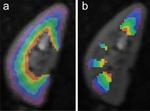
Materials and methods: In this work, we probe the spatial dependence of diffusion MRI metrics with concentrically layered segmentation in 11 healthy kidneys at 3 T. The metrics include those from DTI, IVIM, a combined approach titled "REnal Flow and Microstructure AnisotroPy (REFMAP)", and a multiply encoded model titled "FC-IVIM" providing estimates of fluid velocity and branching length.
Results: Fractional anisotropy decreased from the inner kidney to the outer kidney with the strongest layer correlation in both parenchyma (including cortex and medulla) and medulla with Spearman correlation coefficients and p-values (r, p) of (0.42, <0.001) and (0.37, <0.001), respectively. Also, dynamic parameters derived from the three models significantly decreased with a high correlation from the inner to the outer parenchyma or medulla with (r, p) ranges of (0.46-0.55, <0.001).
Conclusions: These spatial trends might find implications for indirect assessments of kidney physiology and microstructure using diffusion MRI.
期刊介绍:
MAGMA is a multidisciplinary international journal devoted to the publication of articles on all aspects of magnetic resonance techniques and their applications in medicine and biology. MAGMA currently publishes research papers, reviews, letters to the editor, and commentaries, six times a year. The subject areas covered by MAGMA include:
advances in materials, hardware and software in magnetic resonance technology,
new developments and results in research and practical applications of magnetic resonance imaging and spectroscopy related to biology and medicine,
study of animal models and intact cells using magnetic resonance,
reports of clinical trials on humans and clinical validation of magnetic resonance protocols.

 求助内容:
求助内容: 应助结果提醒方式:


