Feasibility and efficacy of endoscopy with blue laser imaging for the detection and diagnosis of cardia polyps: A single-center randomized controlled study
Jia Wei Chen, Gang Liu, Yan Fang Lin, Ting You, Li Jia Yao, Bao Shan Wang, Qing Hua Wang, Da Zhou Li, Wen Wang
{"title":"Feasibility and efficacy of endoscopy with blue laser imaging for the detection and diagnosis of cardia polyps: A single-center randomized controlled study","authors":"Jia Wei Chen, Gang Liu, Yan Fang Lin, Ting You, Li Jia Yao, Bao Shan Wang, Qing Hua Wang, Da Zhou Li, Wen Wang","doi":"10.1111/1751-2980.13267","DOIUrl":null,"url":null,"abstract":"<div>\n \n <section>\n \n <h3> Objective</h3>\n \n <p>To compare the detection rate and diagnostic accuracy of cardia polyps using endoscopy with blue laser imaging (BLI) and white-light imaging (WLI).</p>\n </section>\n \n <section>\n \n <h3> Methods</h3>\n \n <p>Patients were randomly divided into the BLI group and WLI group according to the endoscopic procedures. BLI followed by WLI was conducted in the BLI group, whereas WLI followed by BLI examination was conducted in the WLI group. The number, size, microstructure, and microvascular patterns of cardia polyps detected were recorded. Biopsy of the polyps was then performed.</p>\n </section>\n \n <section>\n \n <h3> Results</h3>\n \n <p>The detection rate of cardia polyps in the BLI group was higher than that in the WLI group (7.87% vs 4.22%, <i>P</i> = 0.018). The rate of overlooked lesions in the BLI group was lower than in the WLI group (0.64% vs 3.38%, <i>P</i> = 0.003). The diagnostic coincidence rate between magnifying BLI and histopathology was 88.16%. The sensitivity, specificity, positive predictive value and negative predictive value for the diagnosis of neoplastic lesions by magnifying endoscopy with BLI were 90.91%, 87.69%, 55.56%, and 98.28%, respectively. The most remarkable patterns for predicting inflammatory polyps were the prolonged and fine network patterns (sensitivity 71.43%, specificity 93.75%). Small round combined with honeycomb patterns were the most common among fundic gland polyps (sensitivity 80.00%, specificity 98.48%). Neoplastic lesions presented as villous or ridge-like combined with core vascular or unclear pattern for both microvascular and microstructure patterns.</p>\n </section>\n \n <section>\n \n <h3> Conclusion</h3>\n \n <p>BLI is more effective than WLI in the detection and diagnosis of cardia polyps, and magnifying endoscopy with BLI may help diagnose such lesions.</p>\n </section>\n </div>","PeriodicalId":15564,"journal":{"name":"Journal of Digestive Diseases","volume":"25 3","pages":"191-199"},"PeriodicalIF":2.3000,"publicationDate":"2024-05-02","publicationTypes":"Journal Article","fieldsOfStudy":null,"isOpenAccess":false,"openAccessPdf":"","citationCount":"0","resultStr":null,"platform":"Semanticscholar","paperid":null,"PeriodicalName":"Journal of Digestive Diseases","FirstCategoryId":"3","ListUrlMain":"https://onlinelibrary.wiley.com/doi/10.1111/1751-2980.13267","RegionNum":3,"RegionCategory":"医学","ArticlePicture":[],"TitleCN":null,"AbstractTextCN":null,"PMCID":null,"EPubDate":"","PubModel":"","JCR":"Q3","JCRName":"GASTROENTEROLOGY & HEPATOLOGY","Score":null,"Total":0}
引用次数: 0
Abstract
Objective
To compare the detection rate and diagnostic accuracy of cardia polyps using endoscopy with blue laser imaging (BLI) and white-light imaging (WLI).
Methods
Patients were randomly divided into the BLI group and WLI group according to the endoscopic procedures. BLI followed by WLI was conducted in the BLI group, whereas WLI followed by BLI examination was conducted in the WLI group. The number, size, microstructure, and microvascular patterns of cardia polyps detected were recorded. Biopsy of the polyps was then performed.
Results
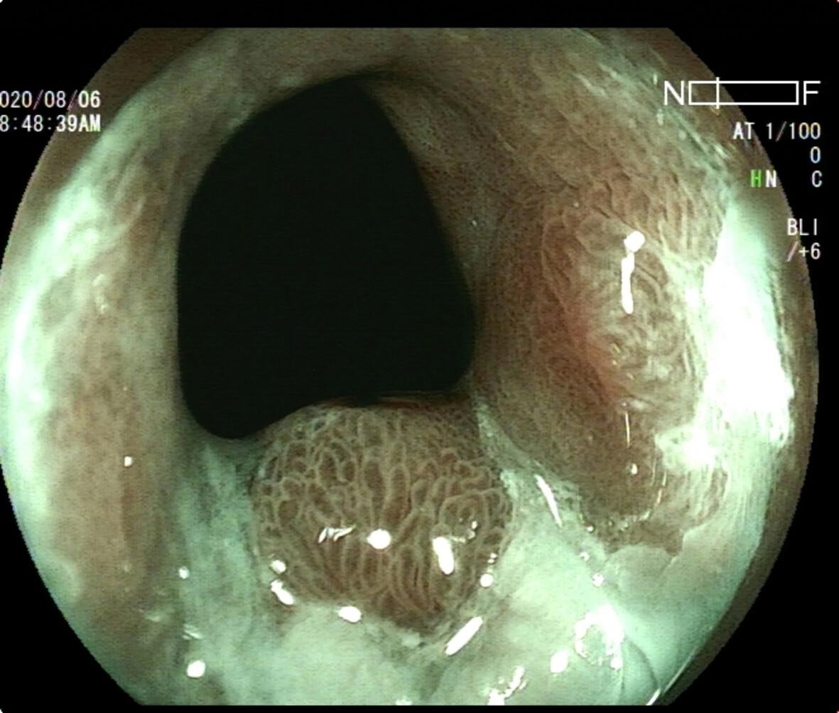
The detection rate of cardia polyps in the BLI group was higher than that in the WLI group (7.87% vs 4.22%, P = 0.018). The rate of overlooked lesions in the BLI group was lower than in the WLI group (0.64% vs 3.38%, P = 0.003). The diagnostic coincidence rate between magnifying BLI and histopathology was 88.16%. The sensitivity, specificity, positive predictive value and negative predictive value for the diagnosis of neoplastic lesions by magnifying endoscopy with BLI were 90.91%, 87.69%, 55.56%, and 98.28%, respectively. The most remarkable patterns for predicting inflammatory polyps were the prolonged and fine network patterns (sensitivity 71.43%, specificity 93.75%). Small round combined with honeycomb patterns were the most common among fundic gland polyps (sensitivity 80.00%, specificity 98.48%). Neoplastic lesions presented as villous or ridge-like combined with core vascular or unclear pattern for both microvascular and microstructure patterns.
Conclusion
BLI is more effective than WLI in the detection and diagnosis of cardia polyps, and magnifying endoscopy with BLI may help diagnose such lesions.
期刊介绍:
The Journal of Digestive Diseases is the official English-language journal of the Chinese Society of Gastroenterology. The journal is published twelve times per year and includes peer-reviewed original papers, review articles and commentaries concerned with research relating to the esophagus, stomach, small intestine, colon, liver, biliary tract and pancreas.
 求助内容:
求助内容: 应助结果提醒方式:


