Characterization and modulation of the pro-inflammatory effects of immune cells in the canine intervertebral disk

IF 3.4 3区 医学 Q1 ORTHOPEDICS
JOR Spine Pub Date : 2024-04-23 DOI:10.1002/jsp2.1333
Mary K. Heimann, Kelly Thompson, Gilian Gunsch, Shirley N. Tang, Brett Klamer, Kara Corps, Benjamin A. Walter, Sarah A. Moore, Devina Purmessur
{"title":"Characterization and modulation of the pro-inflammatory effects of immune cells in the canine intervertebral disk","authors":"Mary K. Heimann,&nbsp;Kelly Thompson,&nbsp;Gilian Gunsch,&nbsp;Shirley N. Tang,&nbsp;Brett Klamer,&nbsp;Kara Corps,&nbsp;Benjamin A. Walter,&nbsp;Sarah A. Moore,&nbsp;Devina Purmessur","doi":"10.1002/jsp2.1333","DOIUrl":null,"url":null,"abstract":"<div>\n \n \n <section>\n \n <h3> Background</h3>\n \n <p>Intervertebral disk (IVD) degeneration affects both humans and canines and is a major cause of low back pain (LBP). Mast cell (MC) and macrophage (MØ) infiltration has been identified in the pathogenesis of IVD degeneration (IVDD) in the human and rodent model but remains understudied in the canine. MC degranulation in the IVD leads to a pro-inflammatory cascade and activates protease activated receptor 2 (PAR2) on IVD cells. The objectives of the present study are to: (1) highlight the pathophysiological changes observed in the degenerate canine IVD, (2) further characterize the inflammatory effect of MCs co-cultured with canine nucleus pulposus (NP) cells, (3) evaluate the effect of construct stiffness on NP and MCs, and (4) identify potential therapeutics to mitigate pathologic changes in the IVD microenvironment.</p>\n </section>\n \n <section>\n \n <h3> Methods</h3>\n \n <p>Canine IVD tissue was isolated from healthy autopsy research dogs (beagle) and pet dogs undergoing laminectomy for IVD herniation. Morphology, protein content, and inflammatory markers were assessed. NP cells isolated from healthy autopsy (Mongrel hounds) tissue were co-cultured with canine MCs within agarose constructs and treated with cromolyn sodium (CS) and PAR2 antagonist (PAR2A). Gene expression, sulfated glycosaminoglycan content, and stiffness of constructs were assessed.</p>\n </section>\n \n <section>\n \n <h3> Results</h3>\n \n <p>CD 31+ blood vessels, mast cell tryptase, and macrophage CD 163+ were increased in the degenerate surgical canine tissue compared to healthy autopsy. Pro-inflammatory genes were upregulated when canine NP cells were co-cultured with MCs and the stiffer microenvironment enhanced these effects. Treatment with CS and PAR2 inhibitors mediated key pro-inflammatory markers in canine NP cells.</p>\n </section>\n \n <section>\n \n <h3> Conclusion</h3>\n \n <p>There is increased MC, MØs, and vascular ingrowth in the degenerate canine IVD tissue, similar to observations in the clinical population with IVDD and LBP. MCs co-cultured with canine NP cells drive inflammation, and CS and PAR2A are potential therapeutics that may mitigate the pathophysiology of IVDD in vitro.</p>\n </section>\n </div>","PeriodicalId":14876,"journal":{"name":"JOR Spine","volume":"7 2","pages":""},"PeriodicalIF":3.4000,"publicationDate":"2024-04-23","publicationTypes":"Journal Article","fieldsOfStudy":null,"isOpenAccess":false,"openAccessPdf":"https://onlinelibrary.wiley.com/doi/epdf/10.1002/jsp2.1333","citationCount":"0","resultStr":null,"platform":"Semanticscholar","paperid":null,"PeriodicalName":"JOR Spine","FirstCategoryId":"3","ListUrlMain":"https://onlinelibrary.wiley.com/doi/10.1002/jsp2.1333","RegionNum":3,"RegionCategory":"医学","ArticlePicture":[],"TitleCN":null,"AbstractTextCN":null,"PMCID":null,"EPubDate":"","PubModel":"","JCR":"Q1","JCRName":"ORTHOPEDICS","Score":null,"Total":0}
引用次数: 0

Abstract

Background

Intervertebral disk (IVD) degeneration affects both humans and canines and is a major cause of low back pain (LBP). Mast cell (MC) and macrophage (MØ) infiltration has been identified in the pathogenesis of IVD degeneration (IVDD) in the human and rodent model but remains understudied in the canine. MC degranulation in the IVD leads to a pro-inflammatory cascade and activates protease activated receptor 2 (PAR2) on IVD cells. The objectives of the present study are to: (1) highlight the pathophysiological changes observed in the degenerate canine IVD, (2) further characterize the inflammatory effect of MCs co-cultured with canine nucleus pulposus (NP) cells, (3) evaluate the effect of construct stiffness on NP and MCs, and (4) identify potential therapeutics to mitigate pathologic changes in the IVD microenvironment.

Methods

Canine IVD tissue was isolated from healthy autopsy research dogs (beagle) and pet dogs undergoing laminectomy for IVD herniation. Morphology, protein content, and inflammatory markers were assessed. NP cells isolated from healthy autopsy (Mongrel hounds) tissue were co-cultured with canine MCs within agarose constructs and treated with cromolyn sodium (CS) and PAR2 antagonist (PAR2A). Gene expression, sulfated glycosaminoglycan content, and stiffness of constructs were assessed.

Results

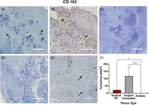
CD 31+ blood vessels, mast cell tryptase, and macrophage CD 163+ were increased in the degenerate surgical canine tissue compared to healthy autopsy. Pro-inflammatory genes were upregulated when canine NP cells were co-cultured with MCs and the stiffer microenvironment enhanced these effects. Treatment with CS and PAR2 inhibitors mediated key pro-inflammatory markers in canine NP cells.

Conclusion

There is increased MC, MØs, and vascular ingrowth in the degenerate canine IVD tissue, similar to observations in the clinical population with IVDD and LBP. MCs co-cultured with canine NP cells drive inflammation, and CS and PAR2A are potential therapeutics that may mitigate the pathophysiology of IVDD in vitro.
Abstract Image

犬椎间盘中免疫细胞促炎作用的特征和调节作用
背景椎间盘(IVD)退化既影响人类也影响犬类,是腰背痛(LBP)的主要原因。已发现肥大细胞(MC)和巨噬细胞(MØ)浸润是人类和啮齿类动物模型中椎间盘退变(IVDD)的发病机制,但对犬的研究仍然不足。IVD 中的 MC 脱颗粒会导致促炎级联反应,并激活 IVD 细胞上的蛋白酶激活受体 2 (PAR2)。本研究的目的是(1)强调在退行性犬 IVD 中观察到的病理生理变化;(2)进一步描述 MCs 与犬髓核 (NP) 细胞共培养的炎症效应;(3)评估构造硬度对 NP 和 MCs 的影响;以及(4)确定缓解 IVD 微环境病理变化的潜在疗法。 方法 从健康的解剖研究犬(小猎犬)和因 IVD 椎间盘突出而接受椎板切除术的宠物犬身上分离犬 IVD 组织。对组织形态、蛋白质含量和炎症标记物进行了评估。从健康尸检犬(猎犬)组织中分离出的 NP 细胞与犬 MCs 共同培养在琼脂糖构建体中,并用色甘酸钠(CS)和 PAR2 拮抗剂(PAR2A)处理。对构建体的基因表达、硫酸化氨基糖含量和硬度进行了评估。 结果 与健康尸检结果相比,变性手术犬组织中的 CD 31+ 血管、肥大细胞胰蛋白酶和巨噬细胞 CD 163+ 均有所增加。当犬 NP 细胞与 MCs 共同培养时,促炎基因上调,而较硬的微环境增强了这些效应。用 CS 和 PAR2 抑制剂处理可介导犬 NP 细胞中的主要促炎标记物。 结论 退行性犬 IVD 组织中 MC、MØs 和血管生长增加,这与 IVDD 和 LBP 临床人群中的观察结果相似。与犬 NP 细胞共同培养的 MCs 会引发炎症,而 CS 和 PAR2A 是潜在的治疗药物,可减轻体外 IVDD 的病理生理学。
本文章由计算机程序翻译,如有差异,请以英文原文为准。
求助全文
约1分钟内获得全文 求助全文
来源期刊
JOR Spine
JOR Spine ORTHOPEDICS-
CiteScore
6.40
自引率
18.90%
发文量
42
审稿时长
10 weeks
×
引用
GB/T 7714-2015
复制
MLA
复制
APA
复制
导出至
BibTeX EndNote RefMan NoteFirst NoteExpress
×
提示
您的信息不完整,为了账户安全,请先补充。
现在去补充
×
提示
您因"违规操作"
具体请查看互助需知
我知道了
×
提示
确定
请完成安全验证×
copy
已复制链接
快去分享给好友吧!
我知道了
右上角分享
点击右上角分享
0
联系我们:info@booksci.cn Book学术提供免费学术资源搜索服务,方便国内外学者检索中英文文献。致力于提供最便捷和优质的服务体验。 Copyright © 2023 布克学术 All rights reserved.
京ICP备2023020795号-1
ghs 京公网安备 11010802042870号
Book学术文献互助
Book学术文献互助群
群 号:481959085
Book学术官方微信