Hidenori Hayashi MD , Jacqueline Contento BSE , Hiroshi Matsushita MD , Paige Mass MS , Vincent Cleveland MS , Seda Aslan MS , Amartya Dave BS , Raquel dos Santos , Angie Zhu , Emmett Reid , Tatsuya Watanabe MD, PhD , Nora Lee MPAP, PA-C , Tyler Dunn BS , Umar Siddiqi , Katherine Nurminsky BS , Vivian Nguyen BA , Keigo Kawaji PhD , Joey Huddle MS , Luka Pocivavsek MD, PhD , Jed Johnson PhD , Narutoshi Hibino MD, PhD
{"title":"Patient-specific tissue engineered vascular graft for aortic arch reconstruction","authors":"Hidenori Hayashi MD , Jacqueline Contento BSE , Hiroshi Matsushita MD , Paige Mass MS , Vincent Cleveland MS , Seda Aslan MS , Amartya Dave BS , Raquel dos Santos , Angie Zhu , Emmett Reid , Tatsuya Watanabe MD, PhD , Nora Lee MPAP, PA-C , Tyler Dunn BS , Umar Siddiqi , Katherine Nurminsky BS , Vivian Nguyen BA , Keigo Kawaji PhD , Joey Huddle MS , Luka Pocivavsek MD, PhD , Jed Johnson PhD , Narutoshi Hibino MD, PhD","doi":"10.1016/j.xjon.2024.02.012","DOIUrl":null,"url":null,"abstract":"<div><h3>Objective(s)</h3><p>The complexity of aortic arch reconstruction due to diverse 3-dimensional geometrical abnormalities is a major challenge. This study introduces 3-dimensional printed tissue-engineered vascular grafts, which can fit patient-specific dimensions, optimize hemodynamics, exhibit antithrombotic and anti-infective properties, and accommodate growth.</p></div><div><h3>Methods</h3><p>We procured cardiac magnetic resonance imaging with 4-dimensional flow for native porcine anatomy (n = 10), from which we designed tissue-engineered vascular grafts for the distal aortic arch, 4 weeks before surgery. An optimal shape of the curved vascular graft was designed using computer-aided design informed by computational fluid dynamics analysis. Grafts were manufactured and implanted into the distal aortic arch of porcine models, and postoperative cardiac magnetic resonance imaging data were collected. Pre- and postimplant hemodynamic data and histology were analyzed.</p></div><div><h3>Results</h3><p>Postoperative magnetic resonance imaging of all pigs with 1:1 ratio of polycaprolactone and poly-L-lactide-co-ε-caprolactone demonstrated no specific dilatation or stenosis of the graft, revealing a positive growth trend in the graft area from the day after surgery to 3 months later, with maintaining a similar shape. The peak wall shear stress of the polycaprolactone/poly-L-lactide-co-ε-caprolactone graft portion did not change significantly between the day after surgery and 3 months later. Immunohistochemistry showed endothelization and smooth muscle layer formation without calcification of the polycaprolactone/poly-L-lactide-co-ε-caprolactone graft.</p></div><div><h3>Conclusions</h3><p>Our patient-specific polycaprolactone/poly-L-lactide-co-ε-caprolactone tissue-engineered vascular grafts demonstrated optimal anatomical fit maintaining ideal hemodynamics and neotissue formation in a porcine model. This study provides a proof of concept of patient-specific tissue-engineered vascular grafts for aortic arch reconstruction.</p></div>","PeriodicalId":74032,"journal":{"name":"JTCVS open","volume":null,"pages":null},"PeriodicalIF":0.0000,"publicationDate":"2024-04-01","publicationTypes":"Journal Article","fieldsOfStudy":null,"isOpenAccess":false,"openAccessPdf":"https://www.sciencedirect.com/science/article/pii/S2666273624000470/pdfft?md5=83578d921ec116ef7b7a1810d2355980&pid=1-s2.0-S2666273624000470-main.pdf","citationCount":"0","resultStr":null,"platform":"Semanticscholar","paperid":null,"PeriodicalName":"JTCVS open","FirstCategoryId":"1085","ListUrlMain":"https://www.sciencedirect.com/science/article/pii/S2666273624000470","RegionNum":0,"RegionCategory":null,"ArticlePicture":[],"TitleCN":null,"AbstractTextCN":null,"PMCID":null,"EPubDate":"","PubModel":"","JCR":"","JCRName":"","Score":null,"Total":0}
引用次数: 0
Abstract
Objective(s)
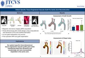
The complexity of aortic arch reconstruction due to diverse 3-dimensional geometrical abnormalities is a major challenge. This study introduces 3-dimensional printed tissue-engineered vascular grafts, which can fit patient-specific dimensions, optimize hemodynamics, exhibit antithrombotic and anti-infective properties, and accommodate growth.
Methods
We procured cardiac magnetic resonance imaging with 4-dimensional flow for native porcine anatomy (n = 10), from which we designed tissue-engineered vascular grafts for the distal aortic arch, 4 weeks before surgery. An optimal shape of the curved vascular graft was designed using computer-aided design informed by computational fluid dynamics analysis. Grafts were manufactured and implanted into the distal aortic arch of porcine models, and postoperative cardiac magnetic resonance imaging data were collected. Pre- and postimplant hemodynamic data and histology were analyzed.
Results
Postoperative magnetic resonance imaging of all pigs with 1:1 ratio of polycaprolactone and poly-L-lactide-co-ε-caprolactone demonstrated no specific dilatation or stenosis of the graft, revealing a positive growth trend in the graft area from the day after surgery to 3 months later, with maintaining a similar shape. The peak wall shear stress of the polycaprolactone/poly-L-lactide-co-ε-caprolactone graft portion did not change significantly between the day after surgery and 3 months later. Immunohistochemistry showed endothelization and smooth muscle layer formation without calcification of the polycaprolactone/poly-L-lactide-co-ε-caprolactone graft.
Conclusions
Our patient-specific polycaprolactone/poly-L-lactide-co-ε-caprolactone tissue-engineered vascular grafts demonstrated optimal anatomical fit maintaining ideal hemodynamics and neotissue formation in a porcine model. This study provides a proof of concept of patient-specific tissue-engineered vascular grafts for aortic arch reconstruction.

 求助内容:
求助内容: 应助结果提醒方式:


