Julie P. Vidal, Lola Danet, Patrice Péran, Jérémie Pariente, Meritxell Bach Cuadra, Natalie M. Zahr, Emmanuel J. Barbeau, Manojkumar Saranathan
{"title":"Robust thalamic nuclei segmentation from T1-weighted MRI using polynomial intensity transformation","authors":"Julie P. Vidal, Lola Danet, Patrice Péran, Jérémie Pariente, Meritxell Bach Cuadra, Natalie M. Zahr, Emmanuel J. Barbeau, Manojkumar Saranathan","doi":"10.1007/s00429-024-02777-5","DOIUrl":null,"url":null,"abstract":"<h3>Abstract</h3> <p>Accurate segmentation of thalamic nuclei, crucial for understanding their role in healthy cognition and in pathologies, is challenging to achieve on standard T1-weighted (T1w) magnetic resonance imaging (MRI) due to poor image contrast. White-matter-nulled (WMn) MRI sequences improve intrathalamic contrast but are not part of clinical protocols or extant databases. In this study, we introduce histogram-based polynomial synthesis (HIPS), a fast preprocessing transform step that synthesizes WMn-like image contrast from standard T1w MRI using a polynomial approximation for intensity transformation. HIPS was incorporated into THalamus Optimized Multi-Atlas Segmentation (THOMAS) pipeline, a method developed and optimized for WMn MRI. HIPS-THOMAS was compared to a convolutional neural network (CNN)-based segmentation method and THOMAS modified for the use of T1w images (T1w-THOMAS). The robustness and accuracy of the three methods were tested across different image contrasts (MPRAGE, SPGR, and MP2RAGE), scanner manufacturers (PHILIPS, GE, and Siemens), and field strengths (3 T and 7 T). HIPS-transformed images improved intra-thalamic contrast and thalamic boundaries, and HIPS-THOMAS yielded significantly higher mean Dice coefficients and reduced volume errors compared to both the CNN method and T1w-THOMAS. Finally, all three methods were compared using the frequently travelling human phantom MRI dataset for inter- and intra-scanner variability, with HIPS displaying the least inter-scanner variability and performing comparably with T1w-THOMAS for intra-scanner variability. In conclusion, our findings highlight the efficacy and robustness of HIPS in enhancing thalamic nuclei segmentation from standard T1w MRI.</p>","PeriodicalId":518000,"journal":{"name":"Brain Structure and Function","volume":"1 1","pages":""},"PeriodicalIF":0.0000,"publicationDate":"2024-03-28","publicationTypes":"Journal Article","fieldsOfStudy":null,"isOpenAccess":false,"openAccessPdf":"","citationCount":"0","resultStr":null,"platform":"Semanticscholar","paperid":null,"PeriodicalName":"Brain Structure and Function","FirstCategoryId":"1085","ListUrlMain":"https://doi.org/10.1007/s00429-024-02777-5","RegionNum":0,"RegionCategory":null,"ArticlePicture":[],"TitleCN":null,"AbstractTextCN":null,"PMCID":null,"EPubDate":"","PubModel":"","JCR":"","JCRName":"","Score":null,"Total":0}
引用次数: 0
Abstract
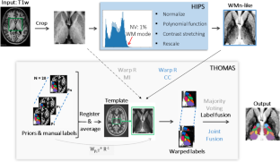
Accurate segmentation of thalamic nuclei, crucial for understanding their role in healthy cognition and in pathologies, is challenging to achieve on standard T1-weighted (T1w) magnetic resonance imaging (MRI) due to poor image contrast. White-matter-nulled (WMn) MRI sequences improve intrathalamic contrast but are not part of clinical protocols or extant databases. In this study, we introduce histogram-based polynomial synthesis (HIPS), a fast preprocessing transform step that synthesizes WMn-like image contrast from standard T1w MRI using a polynomial approximation for intensity transformation. HIPS was incorporated into THalamus Optimized Multi-Atlas Segmentation (THOMAS) pipeline, a method developed and optimized for WMn MRI. HIPS-THOMAS was compared to a convolutional neural network (CNN)-based segmentation method and THOMAS modified for the use of T1w images (T1w-THOMAS). The robustness and accuracy of the three methods were tested across different image contrasts (MPRAGE, SPGR, and MP2RAGE), scanner manufacturers (PHILIPS, GE, and Siemens), and field strengths (3 T and 7 T). HIPS-transformed images improved intra-thalamic contrast and thalamic boundaries, and HIPS-THOMAS yielded significantly higher mean Dice coefficients and reduced volume errors compared to both the CNN method and T1w-THOMAS. Finally, all three methods were compared using the frequently travelling human phantom MRI dataset for inter- and intra-scanner variability, with HIPS displaying the least inter-scanner variability and performing comparably with T1w-THOMAS for intra-scanner variability. In conclusion, our findings highlight the efficacy and robustness of HIPS in enhancing thalamic nuclei segmentation from standard T1w MRI.

 求助内容:
求助内容: 应助结果提醒方式:


