Marie-Helene Ngo , Geraldine S. Pinkus , Eren D. Yeh , Jane E. Brock , Stephanie Schulte , Susan C. Lester
{"title":"Non-sclerosing (T-cell) and sclerosing (B-cell) lymphocytic lobulitis in diagnostic breast biopsies: Clinical, imaging, and pathologic features","authors":"Marie-Helene Ngo , Geraldine S. Pinkus , Eren D. Yeh , Jane E. Brock , Stephanie Schulte , Susan C. Lester","doi":"10.1016/j.humpath.2024.03.006","DOIUrl":null,"url":null,"abstract":"<div><p>Lymphocytic lobulitis (LL) is characterized by prominent lymphocytic infiltrates centered on lobules. Sclerosing lymphocytic lobulitis (SCLL) associated with diabetes mellitus (DM) or autoimmune disease (AI) was the first type to be described. Subsequently, non-sclerosing LL (NSCLL) was reported as an incidental finding in prophylactic mastectomies due to high risk germline mutations or a family history of breast cancer. The two types of LL were distinguished by stromal features and a predominant population of B-cells in the former and T-cells in the latter. In this study, 8 cases of NSCLL detected clinically or by screening were compared to 44 cases of SCLL. One case of NSCLL presented as a palpable mass, 2 as masses on screening, and 5 as MRI enhancement. In contrast, 80% of SCLL cases presented as palpable masses. Half the cases of NSCLL were associated with a <em>BRCA1</em> or <em>2</em> mutation compared to 1 case of SCLL (2%). Three additional cases of NSCLL were associated with a strong family and/or personal history of breast cancer. Almost half (52%) of SCLL cases were associated with DM or AI, but only 25% of NSCLL. Immunoperoxidase studies confirmed a predominance of T-cells in NSCLL and B-cells in SCLL associated with DM or AI. It is important for pathologists to be aware of this new observation that NSCLL can be detected as a palpable mass or an imaging finding in diagnostic biopsies, as its presence can be indicative of a significant risk for breast cancer.</p></div>","PeriodicalId":13062,"journal":{"name":"Human pathology","volume":null,"pages":null},"PeriodicalIF":2.7000,"publicationDate":"2024-03-20","publicationTypes":"Journal Article","fieldsOfStudy":null,"isOpenAccess":false,"openAccessPdf":"https://www.sciencedirect.com/science/article/pii/S0046817724000510/pdfft?md5=7fb4531a979ea6a7ed87df4b5575ea3c&pid=1-s2.0-S0046817724000510-main.pdf","citationCount":"0","resultStr":null,"platform":"Semanticscholar","paperid":null,"PeriodicalName":"Human pathology","FirstCategoryId":"3","ListUrlMain":"https://www.sciencedirect.com/science/article/pii/S0046817724000510","RegionNum":2,"RegionCategory":"医学","ArticlePicture":[],"TitleCN":null,"AbstractTextCN":null,"PMCID":null,"EPubDate":"","PubModel":"","JCR":"Q2","JCRName":"PATHOLOGY","Score":null,"Total":0}
引用次数: 0
Abstract
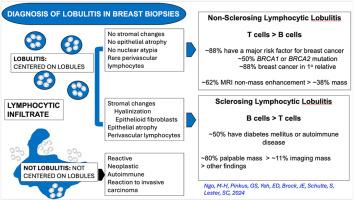
Lymphocytic lobulitis (LL) is characterized by prominent lymphocytic infiltrates centered on lobules. Sclerosing lymphocytic lobulitis (SCLL) associated with diabetes mellitus (DM) or autoimmune disease (AI) was the first type to be described. Subsequently, non-sclerosing LL (NSCLL) was reported as an incidental finding in prophylactic mastectomies due to high risk germline mutations or a family history of breast cancer. The two types of LL were distinguished by stromal features and a predominant population of B-cells in the former and T-cells in the latter. In this study, 8 cases of NSCLL detected clinically or by screening were compared to 44 cases of SCLL. One case of NSCLL presented as a palpable mass, 2 as masses on screening, and 5 as MRI enhancement. In contrast, 80% of SCLL cases presented as palpable masses. Half the cases of NSCLL were associated with a BRCA1 or 2 mutation compared to 1 case of SCLL (2%). Three additional cases of NSCLL were associated with a strong family and/or personal history of breast cancer. Almost half (52%) of SCLL cases were associated with DM or AI, but only 25% of NSCLL. Immunoperoxidase studies confirmed a predominance of T-cells in NSCLL and B-cells in SCLL associated with DM or AI. It is important for pathologists to be aware of this new observation that NSCLL can be detected as a palpable mass or an imaging finding in diagnostic biopsies, as its presence can be indicative of a significant risk for breast cancer.
期刊介绍:
Human Pathology is designed to bring information of clinicopathologic significance to human disease to the laboratory and clinical physician. It presents information drawn from morphologic and clinical laboratory studies with direct relevance to the understanding of human diseases. Papers published concern morphologic and clinicopathologic observations, reviews of diseases, analyses of problems in pathology, significant collections of case material and advances in concepts or techniques of value in the analysis and diagnosis of disease. Theoretical and experimental pathology and molecular biology pertinent to human disease are included. This critical journal is well illustrated with exceptional reproductions of photomicrographs and microscopic anatomy.

 求助内容:
求助内容: 应助结果提醒方式:


