Rodrigo Quevedo García, Sara Arnaiz Díez, Esteban Pérez Pevida, María Lourdes Del Río Solá
{"title":"Orthopantomography Detection of Atheroma Plaques and Its Relationship with Periodontal Disease and Missing Teeth.","authors":"Rodrigo Quevedo García, Sara Arnaiz Díez, Esteban Pérez Pevida, María Lourdes Del Río Solá","doi":"10.1155/2024/8873720","DOIUrl":null,"url":null,"abstract":"<p><strong>Background: </strong>The aim of this study is to determine the atheromatous plaques' prevalence in orthopantomography and their relationship with periodontal disease and missing teeth. <i>Material and Methods</i>. Orthopantomographs of 1,254 patients over 18 years of age from Clínica Arlanza in Lerma, Burgos, were examined between 2017 and 2021. A Planmeca ProOne® orthopantomograph (68 kV, 7 mA, and 10 sg) was used. Statistical analysis was carried out using SPSS Statistics® version 25. The results of the categorical variables were described as frequencies (%). Contingency tables were made with the qualitative variables, and the chi-square test was applied to study the relationship among them. The measure of statistical power used was the relative risk (RR), which was described with its respective 95% confidence interval (CI). Student's <i>t</i>-test was applied to study the relationship between the qualitative variable \"presence or absence of atheroma plaque\" and the quantitative variable \"number of teeth.\"</p><p><strong>Results: </strong>A 6.2% prevalence of atheroma plaques was obtained from 1,079 selected X-rays. The risk in patients with periodontal disease increased as periodontal disease worsened. The risk in patients with periodontal disease increased as periodontal disease worsened as follows: healthy patients vs. periodontal patients with less than 30% bone loss in radiography: RR 0.434, 95% CI 0.181-1.041, <i>p</i> = 0.053 healthy patients vs. patients with between 30%-60% bone loss: RR 0.177, 95% CI 0.075-0.418, <i>p</i> < 0.05 healthy patients vs. patients with more than 60% bone loss: RR 0.121, 95% CI 0.041-0.355, <i>p</i> < 0.05. Patients with calcifications on their orthopantomograms had a lower mean teeth number (20.9 teeth) compared to patients without calcifications (24 teeth), which was statistically significant, <i>t</i> (1077) = -3.125, <i>p</i> < 0.05.</p><p><strong>Conclusions: </strong>Orthopantomography can be considered a screening method to detect patients at increased cardiovascular risk who are referred for individualized study. It is important to continue research to know the real significance of these findings. Dentists should be aware of the importance of our work in our patients' systemic health.</p>","PeriodicalId":51864,"journal":{"name":"Radiology Research and Practice","volume":"2024 ","pages":"8873720"},"PeriodicalIF":1.5000,"publicationDate":"2024-03-04","publicationTypes":"Journal Article","fieldsOfStudy":null,"isOpenAccess":false,"openAccessPdf":"https://www.ncbi.nlm.nih.gov/pmc/articles/PMC10927347/pdf/","citationCount":"0","resultStr":null,"platform":"Semanticscholar","paperid":null,"PeriodicalName":"Radiology Research and Practice","FirstCategoryId":"1085","ListUrlMain":"https://doi.org/10.1155/2024/8873720","RegionNum":0,"RegionCategory":null,"ArticlePicture":[],"TitleCN":null,"AbstractTextCN":null,"PMCID":null,"EPubDate":"2024/1/1 0:00:00","PubModel":"eCollection","JCR":"Q2","JCRName":"RADIOLOGY, NUCLEAR MEDICINE & MEDICAL IMAGING","Score":null,"Total":0}
引用次数: 0
Abstract
Background: The aim of this study is to determine the atheromatous plaques' prevalence in orthopantomography and their relationship with periodontal disease and missing teeth. Material and Methods. Orthopantomographs of 1,254 patients over 18 years of age from Clínica Arlanza in Lerma, Burgos, were examined between 2017 and 2021. A Planmeca ProOne® orthopantomograph (68 kV, 7 mA, and 10 sg) was used. Statistical analysis was carried out using SPSS Statistics® version 25. The results of the categorical variables were described as frequencies (%). Contingency tables were made with the qualitative variables, and the chi-square test was applied to study the relationship among them. The measure of statistical power used was the relative risk (RR), which was described with its respective 95% confidence interval (CI). Student's t-test was applied to study the relationship between the qualitative variable "presence or absence of atheroma plaque" and the quantitative variable "number of teeth."
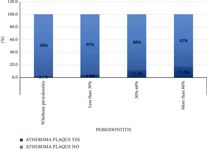
Results: A 6.2% prevalence of atheroma plaques was obtained from 1,079 selected X-rays. The risk in patients with periodontal disease increased as periodontal disease worsened. The risk in patients with periodontal disease increased as periodontal disease worsened as follows: healthy patients vs. periodontal patients with less than 30% bone loss in radiography: RR 0.434, 95% CI 0.181-1.041, p = 0.053 healthy patients vs. patients with between 30%-60% bone loss: RR 0.177, 95% CI 0.075-0.418, p < 0.05 healthy patients vs. patients with more than 60% bone loss: RR 0.121, 95% CI 0.041-0.355, p < 0.05. Patients with calcifications on their orthopantomograms had a lower mean teeth number (20.9 teeth) compared to patients without calcifications (24 teeth), which was statistically significant, t (1077) = -3.125, p < 0.05.
Conclusions: Orthopantomography can be considered a screening method to detect patients at increased cardiovascular risk who are referred for individualized study. It is important to continue research to know the real significance of these findings. Dentists should be aware of the importance of our work in our patients' systemic health.
期刊介绍:
Radiology Research and Practice is a peer-reviewed, Open Access journal that publishes articles on all areas of medical imaging. The journal promotes evidence-based radiology practice though the publication of original research, reviews, and clinical studies for a multidisciplinary audience. Radiology Research and Practice is archived in Portico, which provides permanent archiving for electronic scholarly journals, as well as via the LOCKSS initiative. It operates a fully open access publishing model which allows open global access to its published content. This model is supported through Article Processing Charges. For more information on Article Processing charges in gen

 求助内容:
求助内容: 应助结果提醒方式:


