Accurate detection of cell deformability tracking in hydrodynamic flow by coupling unsupervised and supervised learning

Imen Halima, Mehdi Maleki, Gabriel Frossard, Celine Thomann, Edwin-Joffrey Courtial
{"title":"Accurate detection of cell deformability tracking in hydrodynamic flow by coupling unsupervised and supervised learning","authors":"Imen Halima,&nbsp;Mehdi Maleki,&nbsp;Gabriel Frossard,&nbsp;Celine Thomann,&nbsp;Edwin-Joffrey Courtial","doi":"10.1016/j.mlwa.2024.100538","DOIUrl":null,"url":null,"abstract":"<div><p>The using of deep learning methods in medical images has been successfully used for various applications, including cell segmentation and deformability detection, thereby contributing significantly to advancements in medical analysis. Cell deformability is a fundamental criterion, which must be measured easily and accurately. One common approach for measuring cell deformability is to use microscopy techniques. Recent works have been efforts to develop more advanced and automated methods for measuring cell deformability based on microscopic images, but cell membrane segmentation techniques are still difficult to achieve with precision because of the quality of images. In this paper, we introduce a novel algorithm for cell segmentation that addresses the challenge of microscopic images. AD-MSC cells were controlled by a microfluidic-based system and cell images were acquired by an ultra-fast camera with variable frequency connected to a powerful computer to collect data. The proposed algorithm has a combination of two main components: denoising images using unsupervised learning and cell segmentation and deformability detection using supervised learning which aim to enhance image quality without the need for expensive materials and expert intervention and segment cell deformability with more precision. The contribution of this paper is the combination of two neural networks that treat the database more easily and without the presence of experts. This approach is used to have faster results with high performance according to low datasets from microscopy even with noisy microscopic images. The precision increases to 81 % when we combine DAE with U-Net, compared to 78 % when adding VAE to U-Net and 59 % when using only U-Net.</p></div>","PeriodicalId":74093,"journal":{"name":"Machine learning with applications","volume":"16 ","pages":"Article 100538"},"PeriodicalIF":0.0000,"publicationDate":"2024-02-28","publicationTypes":"Journal Article","fieldsOfStudy":null,"isOpenAccess":false,"openAccessPdf":"https://www.sciencedirect.com/science/article/pii/S2666827024000148/pdfft?md5=1f12d72c76472bd1c4f4bc2e88f648e1&pid=1-s2.0-S2666827024000148-main.pdf","citationCount":"0","resultStr":null,"platform":"Semanticscholar","paperid":null,"PeriodicalName":"Machine learning with applications","FirstCategoryId":"1085","ListUrlMain":"https://www.sciencedirect.com/science/article/pii/S2666827024000148","RegionNum":0,"RegionCategory":null,"ArticlePicture":[],"TitleCN":null,"AbstractTextCN":null,"PMCID":null,"EPubDate":"","PubModel":"","JCR":"","JCRName":"","Score":null,"Total":0}
引用次数: 0

Abstract

The using of deep learning methods in medical images has been successfully used for various applications, including cell segmentation and deformability detection, thereby contributing significantly to advancements in medical analysis. Cell deformability is a fundamental criterion, which must be measured easily and accurately. One common approach for measuring cell deformability is to use microscopy techniques. Recent works have been efforts to develop more advanced and automated methods for measuring cell deformability based on microscopic images, but cell membrane segmentation techniques are still difficult to achieve with precision because of the quality of images. In this paper, we introduce a novel algorithm for cell segmentation that addresses the challenge of microscopic images. AD-MSC cells were controlled by a microfluidic-based system and cell images were acquired by an ultra-fast camera with variable frequency connected to a powerful computer to collect data. The proposed algorithm has a combination of two main components: denoising images using unsupervised learning and cell segmentation and deformability detection using supervised learning which aim to enhance image quality without the need for expensive materials and expert intervention and segment cell deformability with more precision. The contribution of this paper is the combination of two neural networks that treat the database more easily and without the presence of experts. This approach is used to have faster results with high performance according to low datasets from microscopy even with noisy microscopic images. The precision increases to 81 % when we combine DAE with U-Net, compared to 78 % when adding VAE to U-Net and 59 % when using only U-Net.
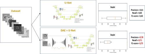
Abstract Image

通过无监督学习和有监督学习的耦合,准确检测流体动力流中的细胞变形性跟踪
在医学图像中使用深度学习方法已成功应用于多种领域,包括细胞分割和可变形性检测,从而极大地推动了医学分析的进步。细胞变形性是一项基本标准,必须能够轻松、准确地测量。测量细胞变形性的一种常见方法是使用显微镜技术。近年来,人们一直在努力开发基于显微图像的更先进、更自动化的细胞变形性测量方法,但由于图像质量的原因,细胞膜分割技术仍难以实现精确测量。在本文中,我们介绍了一种新型的细胞膜分割算法,以应对显微图像带来的挑战。AD-MSC细胞由基于微流控的系统控制,细胞图像由连接到功能强大的计算机的可变频率超快相机采集。所提出的算法由两个主要部分组成:利用无监督学习对图像进行去噪,以及利用有监督学习对细胞进行分割和变形检测,目的是在无需昂贵材料和专家干预的情况下提高图像质量,并更精确地分割细胞变形。本文的贡献在于将两个神经网络结合起来,在没有专家在场的情况下更轻松地处理数据库。根据显微镜的低数据集,即使是嘈杂的显微图像,这种方法也能获得更快的结果和更高的性能。当我们将 DAE 与 U-Net 相结合时,精确度提高到 81%,而将 VAE 添加到 U-Net 时为 78%,仅使用 U-Net 时为 59%。
本文章由计算机程序翻译,如有差异,请以英文原文为准。
求助全文
约1分钟内获得全文 求助全文
来源期刊
Machine learning with applications
Machine learning with applications Management Science and Operations Research, Artificial Intelligence, Computer Science Applications
自引率
0.00%
发文量
0
审稿时长
98 days
×
引用
GB/T 7714-2015
复制
MLA
复制
APA
复制
导出至
BibTeX EndNote RefMan NoteFirst NoteExpress
×
提示
您的信息不完整,为了账户安全,请先补充。
现在去补充
×
提示
您因"违规操作"
具体请查看互助需知
我知道了
×
提示
确定
请完成安全验证×
copy
已复制链接
快去分享给好友吧!
我知道了
右上角分享
点击右上角分享
0
联系我们:info@booksci.cn Book学术提供免费学术资源搜索服务,方便国内外学者检索中英文文献。致力于提供最便捷和优质的服务体验。 Copyright © 2023 布克学术 All rights reserved.
京ICP备2023020795号-1
ghs 京公网安备 11010802042870号
Book学术文献互助
Book学术文献互助群
群 号:481959085
Book学术官方微信