Growth of the brachial nerve plexus with reference to topographical relation of the medianus nerve ansa with the thoracic wall and shoulder: a histologic study using human embryos and fetuses
Kwang Ho Cho, Ji Hyun Kim, Masahito Yamamoto, Shogo Hayashi, Gen Murakami, Jose Francisco Rodríguez-Vázquez
{"title":"Growth of the brachial nerve plexus with reference to topographical relation of the medianus nerve ansa with the thoracic wall and shoulder: a histologic study using human embryos and fetuses","authors":"Kwang Ho Cho, Ji Hyun Kim, Masahito Yamamoto, Shogo Hayashi, Gen Murakami, Jose Francisco Rodríguez-Vázquez","doi":"10.1007/s00276-024-03317-w","DOIUrl":null,"url":null,"abstract":"<h3 data-test=\"abstract-sub-heading\">Background</h3><p>There is currently no information on positional changes in the brachial nerve plexus during prenatal growth. The subclavian–axillary artery passing through the medianus nerve ansa is considered a good landmark for evaluating the height of the plexus.</p><h3 data-test=\"abstract-sub-heading\">Materials and methods</h3><p>We used histologic sections from 9 embryos and 17 fetuses (approximately 6–15 weeks of gestational age) to identify the height of the ansa by referring to the level of the rib and the glenohumeral joint.</p><h3 data-test=\"abstract-sub-heading\">Results</h3><p>The nerve ansa was usually (23 plexuses) observed at the level of the first and/or second ribs. However, it was sometimes observed above the first rib, at a distance equal to or more than an intercostal width (7 plexuses). In the latter group, the ansa was usually located below the glenohumeral joint. Thus, the joint was located higher than the first rib, although the upper extremities were in the anatomic position for all specimens. The left–right difference in the height of the plexus corresponded to or was less than the width of the first intercostal space. Despite the synchronized growth between the thorax and shoulder girdle, the brachial plexus showed a considerable variation in comparative height; the range corresponded to twice of an intercostal width. Whether the nerve plexus is located high or low is determined at an early developmental stage and is maintained during the later growth stages.</p><h3 data-test=\"abstract-sub-heading\">Conclusion</h3><p>The high-positioned plexus might cause nerve injury at delivery, followed by a glenohumeral joint deformity because of the fragility without fixation in the thorax.</p>","PeriodicalId":49296,"journal":{"name":"Surgical and Radiologic Anatomy","volume":"50 1","pages":""},"PeriodicalIF":1.2000,"publicationDate":"2024-03-03","publicationTypes":"Journal Article","fieldsOfStudy":null,"isOpenAccess":false,"openAccessPdf":"","citationCount":"0","resultStr":null,"platform":"Semanticscholar","paperid":null,"PeriodicalName":"Surgical and Radiologic Anatomy","FirstCategoryId":"3","ListUrlMain":"https://doi.org/10.1007/s00276-024-03317-w","RegionNum":4,"RegionCategory":"医学","ArticlePicture":[],"TitleCN":null,"AbstractTextCN":null,"PMCID":null,"EPubDate":"","PubModel":"","JCR":"Q3","JCRName":"ANATOMY & MORPHOLOGY","Score":null,"Total":0}
引用次数: 0
Abstract
Background
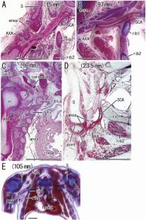
There is currently no information on positional changes in the brachial nerve plexus during prenatal growth. The subclavian–axillary artery passing through the medianus nerve ansa is considered a good landmark for evaluating the height of the plexus.
Materials and methods
We used histologic sections from 9 embryos and 17 fetuses (approximately 6–15 weeks of gestational age) to identify the height of the ansa by referring to the level of the rib and the glenohumeral joint.
Results
The nerve ansa was usually (23 plexuses) observed at the level of the first and/or second ribs. However, it was sometimes observed above the first rib, at a distance equal to or more than an intercostal width (7 plexuses). In the latter group, the ansa was usually located below the glenohumeral joint. Thus, the joint was located higher than the first rib, although the upper extremities were in the anatomic position for all specimens. The left–right difference in the height of the plexus corresponded to or was less than the width of the first intercostal space. Despite the synchronized growth between the thorax and shoulder girdle, the brachial plexus showed a considerable variation in comparative height; the range corresponded to twice of an intercostal width. Whether the nerve plexus is located high or low is determined at an early developmental stage and is maintained during the later growth stages.
Conclusion
The high-positioned plexus might cause nerve injury at delivery, followed by a glenohumeral joint deformity because of the fragility without fixation in the thorax.
期刊介绍:
Anatomy is a morphological science which cannot fail to interest the clinician. The practical application of anatomical research to clinical problems necessitates special adaptation and selectivity in choosing from numerous international works. Although there is a tendency to believe that meaningful advances in anatomy are unlikely, constant revision is necessary. Surgical and Radiologic Anatomy, the first international journal of Clinical anatomy has been created in this spirit.
Its goal is to serve clinicians, regardless of speciality-physicians, surgeons, radiologists or other specialists-as an indispensable aid with which they can improve their knowledge of anatomy. Each issue includes: Original papers, review articles, articles on the anatomical bases of medical, surgical and radiological techniques, articles of normal radiologic anatomy, brief reviews of anatomical publications of clinical interest.
Particular attention is given to high quality illustrations, which are indispensable for a better understanding of anatomical problems.
Surgical and Radiologic Anatomy is a journal written by anatomists for clinicians with a special interest in anatomy.

 求助内容:
求助内容: 应助结果提醒方式:


