Low-intensity pulsed ultrasound induces proliferation of human neural stem cells

Arwa A. Al-Maswary, A. Damien Walmsley, Paul R. Cooper, Ben A. Scheven
{"title":"Low-intensity pulsed ultrasound induces proliferation of human neural stem cells","authors":"Arwa A. Al-Maswary,&nbsp;A. Damien Walmsley,&nbsp;Paul R. Cooper,&nbsp;Ben A. Scheven","doi":"10.1002/ctd2.259","DOIUrl":null,"url":null,"abstract":"<div>\n \n \n <section>\n \n <h3> Background</h3>\n \n <p>Low-intensity pulsed ultrasound (LIPUS) has been highlighted as a potential therapy for tissue repair and regeneration. However, little is known about LIPUS effects on neuromodulation. This research was conducted to study LIPUS effect on the proliferation of human neural stem cells.</p>\n </section>\n \n <section>\n \n <h3> Materials and methods</h3>\n \n <p>The human SH-SY5Y neuroblastoma cell line was used as a neural stem cell model. The well-documented SH-SY5Y neurogenic protocol, which involves treatment with all trans-retinoic acid (ATRA) for 5 days and then brain-derived neurotrophic factor (BDNF) for 7 days, was used to synchronise the growth cell cycle to G1 phase of the cell cycle before proliferation testing. Subsequently, the neural stem cells were then treated with single or triple 20-min LIPUS exposures (Intensity I<sub>SATA</sub>: 60 mW/cm<sup>2</sup>, frequency: 1.5 MHz, pulse repetition: 100 Hz, and duty cycle: 20%). Cell proliferation was analysed using cell counting of β-tubulin and neurofilament medium-positive neural stem cells, Ki67-cell proliferation marker and metabolic-based assays (cell counting kit-8 and alamarBlue). The involvement of ERK signalling was investigated by quantification of phospho-ERK1/2 levels and cell proliferation with and without MEK/ERK inhibitor (U0126).</p>\n </section>\n \n <section>\n \n <h3> Results</h3>\n \n <p>The results show that LIPUS exposure(s) induced cell proliferation, as evidenced by an increase in the numbers of neural stem cells. ERK signalling is involved in LIPUS-induced neural stem cell proliferation, as evidenced by concurrent inhibition of LIPUS-induced phospho-ERK levels and cell proliferation in the presence of the MEK/ERK inhibitor.</p>\n </section>\n \n <section>\n \n <h3> Conclusion</h3>\n \n <p>This study provides original evidence that LIPUS can stimulate neural stem/progenitor cell proliferation. LIPUS may be suggested as a sole or an adjunct therapeutic application to promote the neural stem cell pool in stem cell therapies and tissue engineering approaches for nerve repair and regeneration for the management of traumatic nerve injuries and regenerative endodontic treatment.</p>\n </section>\n </div>","PeriodicalId":72605,"journal":{"name":"Clinical and translational discovery","volume":"4 2","pages":""},"PeriodicalIF":0.0000,"publicationDate":"2024-02-29","publicationTypes":"Journal Article","fieldsOfStudy":null,"isOpenAccess":false,"openAccessPdf":"https://onlinelibrary.wiley.com/doi/epdf/10.1002/ctd2.259","citationCount":"0","resultStr":null,"platform":"Semanticscholar","paperid":null,"PeriodicalName":"Clinical and translational discovery","FirstCategoryId":"1085","ListUrlMain":"https://onlinelibrary.wiley.com/doi/10.1002/ctd2.259","RegionNum":0,"RegionCategory":null,"ArticlePicture":[],"TitleCN":null,"AbstractTextCN":null,"PMCID":null,"EPubDate":"","PubModel":"","JCR":"","JCRName":"","Score":null,"Total":0}
引用次数: 0

Abstract

Background

Low-intensity pulsed ultrasound (LIPUS) has been highlighted as a potential therapy for tissue repair and regeneration. However, little is known about LIPUS effects on neuromodulation. This research was conducted to study LIPUS effect on the proliferation of human neural stem cells.

Materials and methods

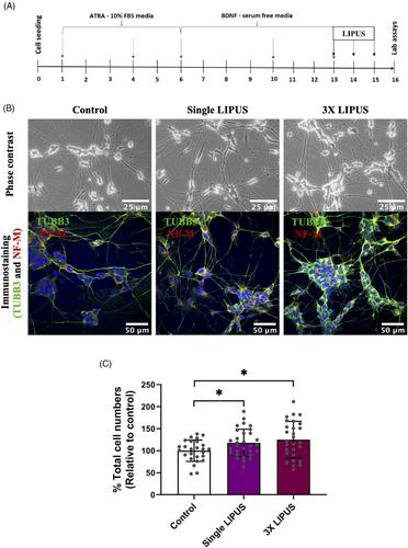
The human SH-SY5Y neuroblastoma cell line was used as a neural stem cell model. The well-documented SH-SY5Y neurogenic protocol, which involves treatment with all trans-retinoic acid (ATRA) for 5 days and then brain-derived neurotrophic factor (BDNF) for 7 days, was used to synchronise the growth cell cycle to G1 phase of the cell cycle before proliferation testing. Subsequently, the neural stem cells were then treated with single or triple 20-min LIPUS exposures (Intensity ISATA: 60 mW/cm2, frequency: 1.5 MHz, pulse repetition: 100 Hz, and duty cycle: 20%). Cell proliferation was analysed using cell counting of β-tubulin and neurofilament medium-positive neural stem cells, Ki67-cell proliferation marker and metabolic-based assays (cell counting kit-8 and alamarBlue). The involvement of ERK signalling was investigated by quantification of phospho-ERK1/2 levels and cell proliferation with and without MEK/ERK inhibitor (U0126).

Results

The results show that LIPUS exposure(s) induced cell proliferation, as evidenced by an increase in the numbers of neural stem cells. ERK signalling is involved in LIPUS-induced neural stem cell proliferation, as evidenced by concurrent inhibition of LIPUS-induced phospho-ERK levels and cell proliferation in the presence of the MEK/ERK inhibitor.

Conclusion

This study provides original evidence that LIPUS can stimulate neural stem/progenitor cell proliferation. LIPUS may be suggested as a sole or an adjunct therapeutic application to promote the neural stem cell pool in stem cell therapies and tissue engineering approaches for nerve repair and regeneration for the management of traumatic nerve injuries and regenerative endodontic treatment.
Abstract Image

低强度脉冲超声诱导人类神经干细胞增殖
背景低强度脉冲超声(LIPUS)作为一种潜在的组织修复和再生疗法已受到重视。然而,人们对 LIPUS 对神经调节的影响知之甚少。本研究旨在研究 LIPUS 对人类神经干细胞增殖的影响。 材料与方法 以人类 SH-SY5Y 神经母细胞瘤细胞系作为神经干细胞模型。SH-SY5Y神经干细胞增殖试验采用的是有据可查的SH-SY5Y神经干细胞增殖方案,即用全反式维甲酸(ATRA)处理5天,然后用脑源性神经营养因子(BDNF)处理7天,在增殖试验前将生长细胞周期同步到细胞周期的G1期。随后,对神经干细胞进行单次或三次 20 分钟的 LIPUS 曝光(ISATA 强度:60 mW/cm2,频率:1.5 MHz,脉冲重复频率:100 Hz,占空比:1.5 MHz):100赫兹,占空比:20%)。细胞增殖分析采用了β-微管蛋白和神经丝介质阳性神经干细胞的细胞计数、Ki67-细胞增殖标记和基于代谢的检测方法(细胞计数试剂盒-8和α蓝)。在使用或不使用 MEK/ERK 抑制剂(U0126)的情况下,通过量化磷酸-ERK1/2 水平和细胞增殖来研究 ERK 信号的参与情况。 结果 结果表明,LIPUS 暴露可诱导细胞增殖,神经干细胞数量的增加就是证明。ERK信号参与了LIPUS诱导的神经干细胞增殖,在MEK/ERK抑制剂存在的情况下,LIPUS诱导的磷酸化ERK水平和细胞增殖同时受到抑制就证明了这一点。 结论 本研究提供了 LIPUS 可刺激神经干细胞/祖细胞增殖的原始证据。在干细胞疗法和组织工程方法中,LIPUS 可作为一种单独或辅助的治疗应用,促进神经干细胞池的神经修复和再生,以治疗创伤性神经损伤和牙髓再生治疗。
本文章由计算机程序翻译,如有差异,请以英文原文为准。
求助全文
约1分钟内获得全文 求助全文
来源期刊
CiteScore
1.00
自引率
0.00%
发文量
0
×
引用
GB/T 7714-2015
复制
MLA
复制
APA
复制
导出至
BibTeX EndNote RefMan NoteFirst NoteExpress
×
提示
您的信息不完整,为了账户安全,请先补充。
现在去补充
×
提示
您因"违规操作"
具体请查看互助需知
我知道了
×
提示
确定
请完成安全验证×
copy
已复制链接
快去分享给好友吧!
我知道了
右上角分享
点击右上角分享
0
联系我们:info@booksci.cn Book学术提供免费学术资源搜索服务,方便国内外学者检索中英文文献。致力于提供最便捷和优质的服务体验。 Copyright © 2023 布克学术 All rights reserved.
京ICP备2023020795号-1
ghs 京公网安备 11010802042870号
Book学术文献互助
Book学术文献互助群
群 号:481959085
Book学术官方微信