Computational methods for metastasis detection in lymph nodes and characterization of the metastasis-free lymph node microarchitecture: A systematic-narrative hybrid review
Elzbieta Budginaite , Derek R. Magee , Maximilian Kloft , Henry C. Woodruff , Heike I. Grabsch
{"title":"Computational methods for metastasis detection in lymph nodes and characterization of the metastasis-free lymph node microarchitecture: A systematic-narrative hybrid review","authors":"Elzbieta Budginaite , Derek R. Magee , Maximilian Kloft , Henry C. Woodruff , Heike I. Grabsch","doi":"10.1016/j.jpi.2024.100367","DOIUrl":null,"url":null,"abstract":"<div><h3>Background</h3><p>Histological examination of tumor draining lymph nodes (LNs) plays a vital role in cancer staging and prognostication. However, as soon as a LN is classed as metastasis-free, no further investigation will be performed and thus, potentially clinically relevant information detectable in tumor-free LNs is currently not captured.</p></div><div><h3>Objective</h3><p>To systematically study and critically assess methods for the analysis of digitized histological LN images described in published research.</p></div><div><h3>Methods</h3><p>A systematic search was conducted in several public databases up to December 2023 using relevant search terms. Studies using brightfield light microscopy images of hematoxylin and eosin or immunohistochemically stained LN tissue sections aiming to detect and/or segment LNs, their compartments or metastatic tumor using artificial intelligence (AI) were included. Dataset, AI methodology, cancer type, and study objective were compared between articles.</p></div><div><h3>Results</h3><p>A total of 7201 articles were collected and 73 articles remained for detailed analyses after article screening. Of the remaining articles, 86% aimed at LN metastasis identification, 8% aimed at LN compartment segmentation, and remaining focused on LN contouring. Furthermore, 78% of articles used patch classification and 22% used pixel segmentation models for analyses. Five out of six studies (83%) of metastasis-free LNs were performed on publicly unavailable datasets, making quantitative article comparison impossible.</p></div><div><h3>Conclusions</h3><p>Multi-scale models mimicking multiple microscopy zooms show promise for computational LN analysis. Large-scale datasets are needed to establish the clinical relevance of analyzing metastasis-free LN in detail. Further research is needed to identify clinically interpretable metrics for LN compartment characterization.</p></div>","PeriodicalId":37769,"journal":{"name":"Journal of Pathology Informatics","volume":"15 ","pages":"Article 100367"},"PeriodicalIF":0.0000,"publicationDate":"2024-02-04","publicationTypes":"Journal Article","fieldsOfStudy":null,"isOpenAccess":false,"openAccessPdf":"https://www.sciencedirect.com/science/article/pii/S2153353924000063/pdfft?md5=d843e63269692c17cbcf96f14d687dad&pid=1-s2.0-S2153353924000063-main.pdf","citationCount":"0","resultStr":null,"platform":"Semanticscholar","paperid":null,"PeriodicalName":"Journal of Pathology Informatics","FirstCategoryId":"1085","ListUrlMain":"https://www.sciencedirect.com/science/article/pii/S2153353924000063","RegionNum":0,"RegionCategory":null,"ArticlePicture":[],"TitleCN":null,"AbstractTextCN":null,"PMCID":null,"EPubDate":"","PubModel":"","JCR":"Q2","JCRName":"Medicine","Score":null,"Total":0}
引用次数: 0
Abstract
Background
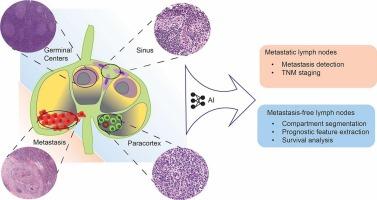
Histological examination of tumor draining lymph nodes (LNs) plays a vital role in cancer staging and prognostication. However, as soon as a LN is classed as metastasis-free, no further investigation will be performed and thus, potentially clinically relevant information detectable in tumor-free LNs is currently not captured.
Objective
To systematically study and critically assess methods for the analysis of digitized histological LN images described in published research.
Methods
A systematic search was conducted in several public databases up to December 2023 using relevant search terms. Studies using brightfield light microscopy images of hematoxylin and eosin or immunohistochemically stained LN tissue sections aiming to detect and/or segment LNs, their compartments or metastatic tumor using artificial intelligence (AI) were included. Dataset, AI methodology, cancer type, and study objective were compared between articles.
Results
A total of 7201 articles were collected and 73 articles remained for detailed analyses after article screening. Of the remaining articles, 86% aimed at LN metastasis identification, 8% aimed at LN compartment segmentation, and remaining focused on LN contouring. Furthermore, 78% of articles used patch classification and 22% used pixel segmentation models for analyses. Five out of six studies (83%) of metastasis-free LNs were performed on publicly unavailable datasets, making quantitative article comparison impossible.
Conclusions
Multi-scale models mimicking multiple microscopy zooms show promise for computational LN analysis. Large-scale datasets are needed to establish the clinical relevance of analyzing metastasis-free LN in detail. Further research is needed to identify clinically interpretable metrics for LN compartment characterization.
期刊介绍:
The Journal of Pathology Informatics (JPI) is an open access peer-reviewed journal dedicated to the advancement of pathology informatics. This is the official journal of the Association for Pathology Informatics (API). The journal aims to publish broadly about pathology informatics and freely disseminate all articles worldwide. This journal is of interest to pathologists, informaticians, academics, researchers, health IT specialists, information officers, IT staff, vendors, and anyone with an interest in informatics. We encourage submissions from anyone with an interest in the field of pathology informatics. We publish all types of papers related to pathology informatics including original research articles, technical notes, reviews, viewpoints, commentaries, editorials, symposia, meeting abstracts, book reviews, and correspondence to the editors. All submissions are subject to rigorous peer review by the well-regarded editorial board and by expert referees in appropriate specialties.

 求助内容:
求助内容: 应助结果提醒方式:


