Mónica Ferreira, Tamara Schaprian, David Kügler, Martin Reuter, Katharina Deike-Hoffmann, Dagmar Timmann, Thomas M Ernst, Paola Giunti, Hector Garcia-Moreno, Bart van de Warrenburg, Judith van Gaalen, Jeroen de Vries, Heike Jacobi, Katharina Marie Steiner, Gülin Öz, James M Joers, Chiadi Onyike, Michal Povazan, Kathrin Reetz, Sandro Romanzetti, Thomas Klockgether, Jennifer Faber
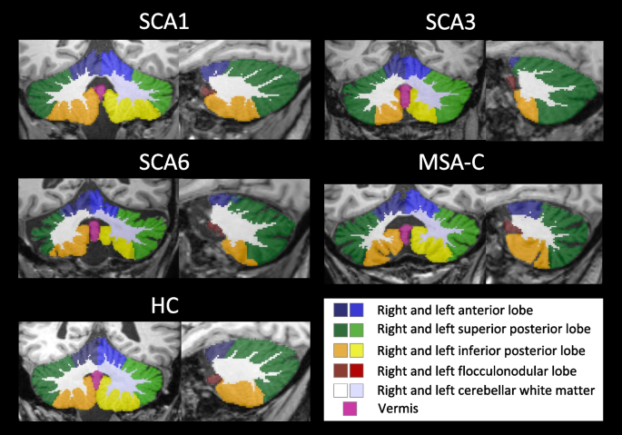
{"title":"Cerebellar Volumetry in Ataxias: Relation to Ataxia Severity and Duration.","authors":"Mónica Ferreira, Tamara Schaprian, David Kügler, Martin Reuter, Katharina Deike-Hoffmann, Dagmar Timmann, Thomas M Ernst, Paola Giunti, Hector Garcia-Moreno, Bart van de Warrenburg, Judith van Gaalen, Jeroen de Vries, Heike Jacobi, Katharina Marie Steiner, Gülin Öz, James M Joers, Chiadi Onyike, Michal Povazan, Kathrin Reetz, Sandro Romanzetti, Thomas Klockgether, Jennifer Faber","doi":"10.1007/s12311-024-01659-0","DOIUrl":null,"url":null,"abstract":"<p><p>Cerebellar atrophy is the neuropathological hallmark of most ataxias. Hence, quantifying the volume of the cerebellar grey and white matter is of great interest. In this study, we aim to identify volume differences in the cerebellum between spinocerebellar ataxia type 1 (SCA1), SCA3 and SCA6 as well as multiple system atrophy of cerebellar type (MSA-C). Our cross-sectional data set comprised mutation carriers of SCA1 (N=12), SCA3 (N=62), SCA6 (N=14), as well as MSA-C patients (N=16). Cerebellar volumes were obtained from T1-weighted magnetic resonance images. To compare the different atrophy patterns, we performed a z-transformation and plotted the intercept of each patient group's model at the mean of 7 years of ataxia duration as well as at the mean ataxia severity of 14 points in the SARA sum score. In addition, we plotted the extrapolation at ataxia duration of 0 years as well as 0 points in the SARA sum score. Patients with MSA-C demonstrated the most pronounced volume loss, particularly in the cerebellar white matter, at the late time intercept. Patients with SCA6 showed a pronounced volume loss in cerebellar grey matter with increasing ataxia severity compared to all other patient groups. MSA-C, SCA1 and SCA3 showed a prominent atrophy of the cerebellar white matter. Our results (i) confirmed SCA6 being considered as a pure cerebellar grey matter disease, (ii) emphasise the involvement of cerebellar white matter in the neuropathology of SCA1, SCA3 and MSA-C, and (iii) reflect the rapid clinical progression in MSA-C.</p>","PeriodicalId":50706,"journal":{"name":"Cerebellum","volume":null,"pages":null},"PeriodicalIF":2.7000,"publicationDate":"2024-08-01","publicationTypes":"Journal Article","fieldsOfStudy":null,"isOpenAccess":false,"openAccessPdf":"https://www.ncbi.nlm.nih.gov/pmc/articles/PMC11269395/pdf/","citationCount":"0","resultStr":null,"platform":"Semanticscholar","paperid":null,"PeriodicalName":"Cerebellum","FirstCategoryId":"3","ListUrlMain":"https://doi.org/10.1007/s12311-024-01659-0","RegionNum":3,"RegionCategory":"医学","ArticlePicture":[],"TitleCN":null,"AbstractTextCN":null,"PMCID":null,"EPubDate":"2024/2/16 0:00:00","PubModel":"Epub","JCR":"Q3","JCRName":"NEUROSCIENCES","Score":null,"Total":0}
引用次数: 0
Abstract
Cerebellar atrophy is the neuropathological hallmark of most ataxias. Hence, quantifying the volume of the cerebellar grey and white matter is of great interest. In this study, we aim to identify volume differences in the cerebellum between spinocerebellar ataxia type 1 (SCA1), SCA3 and SCA6 as well as multiple system atrophy of cerebellar type (MSA-C). Our cross-sectional data set comprised mutation carriers of SCA1 (N=12), SCA3 (N=62), SCA6 (N=14), as well as MSA-C patients (N=16). Cerebellar volumes were obtained from T1-weighted magnetic resonance images. To compare the different atrophy patterns, we performed a z-transformation and plotted the intercept of each patient group's model at the mean of 7 years of ataxia duration as well as at the mean ataxia severity of 14 points in the SARA sum score. In addition, we plotted the extrapolation at ataxia duration of 0 years as well as 0 points in the SARA sum score. Patients with MSA-C demonstrated the most pronounced volume loss, particularly in the cerebellar white matter, at the late time intercept. Patients with SCA6 showed a pronounced volume loss in cerebellar grey matter with increasing ataxia severity compared to all other patient groups. MSA-C, SCA1 and SCA3 showed a prominent atrophy of the cerebellar white matter. Our results (i) confirmed SCA6 being considered as a pure cerebellar grey matter disease, (ii) emphasise the involvement of cerebellar white matter in the neuropathology of SCA1, SCA3 and MSA-C, and (iii) reflect the rapid clinical progression in MSA-C.
期刊介绍:
Official publication of the Society for Research on the Cerebellum devoted to genetics of cerebellar ataxias, role of cerebellum in motor control and cognitive function, and amid an ageing population, diseases associated with cerebellar dysfunction.
The Cerebellum is a central source for the latest developments in fundamental neurosciences including molecular and cellular biology; behavioural neurosciences and neurochemistry; genetics; fundamental and clinical neurophysiology; neurology and neuropathology; cognition and neuroimaging.
The Cerebellum benefits neuroscientists in molecular and cellular biology; neurophysiologists; researchers in neurotransmission; neurologists; radiologists; paediatricians; neuropsychologists; students of neurology and psychiatry and others.
 求助内容:
求助内容: 应助结果提醒方式:


