{"title":"A case of low-grade endometrial stromal sarcoma presented as an intramyometrial mass mimicking uterine leiomyoma on MRI.","authors":"Soichiro Tamada, Hiromi Edo, Taishi Sakima, Ryo Tanaka, Kohei Shikata, Soko Nishitani, Morikazu Miyamoto, Masashi Takano, Keisuke Kuboshima, Kosuke Miyai, Sho Ogata, Hiroshi Shinmoto","doi":"10.1093/bjrcr/uaad012","DOIUrl":null,"url":null,"abstract":"<p><p>A low-grade endometrial stromal sarcoma (ESS) has a pattern of presenting as an intramyometrial mass and is often misdiagnosed as cellular leiomyoma or degenerative uterine leiomyoma. A low-grade ESS is a malignant tumour that requires total hysterectomy with bilateral salpingo-oophorectomy; while a leiomyoma is a benign tumour and could be acceptable for enucleation. As the treatment strategies differ between a low-grade ESS and leiomyoma, radiologists should be familiar with the characteristic MRI findings of a low-grade ESS. A 51-year-old woman with abnormal uterine bleeding had been observed for 2 years at a previous hospital for a uterine leiomyoma based on MRI findings. A contrast-enhanced MRI demonstrated an intramyometrial mass composed of three components with the hypointense rim on T2-weighted images (T2WI): the first component was a homogeneous solid structure with mild hyperintensity on T2WI with a low apparent diffusion coefficient value; the second component was cystic; the third component was a structure of low signal intensity on T2WI similar to the muscle. Although a degenerative uterine leiomyoma was a differential diagnosis, these MRI findings were suggestive of a low-grade ESS. A total abdominal hysterectomy, bilateral salpingo-oophorectomy, pelvic lymphadenectomy, and partial omentectomy were performed. The pathological diagnosis was a low-grade ESS. In a low-grade ESS, there are three major patterns of MRI findings: one of these patterns is the less popular but clinically important intramyometrial mass pattern, which can be misdiagnosed as a leiomyoma, and this case conformed to this pattern.</p>","PeriodicalId":45216,"journal":{"name":"BJR Case Reports","volume":"10 1","pages":"uaad012"},"PeriodicalIF":0.5000,"publicationDate":"2023-12-18","publicationTypes":"Journal Article","fieldsOfStudy":null,"isOpenAccess":false,"openAccessPdf":"https://www.ncbi.nlm.nih.gov/pmc/articles/PMC10860526/pdf/","citationCount":"0","resultStr":null,"platform":"Semanticscholar","paperid":null,"PeriodicalName":"BJR Case Reports","FirstCategoryId":"1085","ListUrlMain":"https://doi.org/10.1093/bjrcr/uaad012","RegionNum":0,"RegionCategory":null,"ArticlePicture":[],"TitleCN":null,"AbstractTextCN":null,"PMCID":null,"EPubDate":"2024/1/1 0:00:00","PubModel":"eCollection","JCR":"Q4","JCRName":"RADIOLOGY, NUCLEAR MEDICINE & MEDICAL IMAGING","Score":null,"Total":0}
引用次数: 0
Abstract
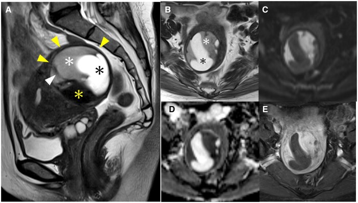
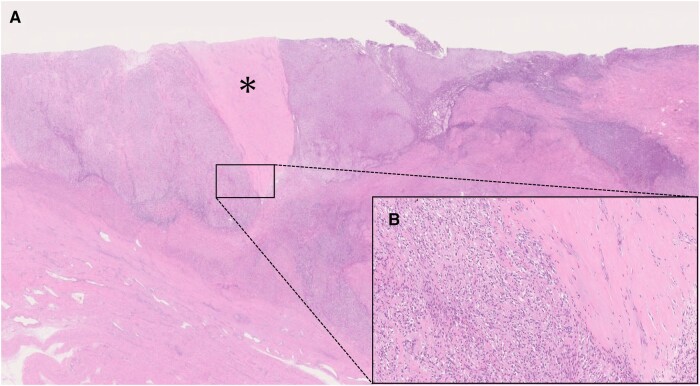
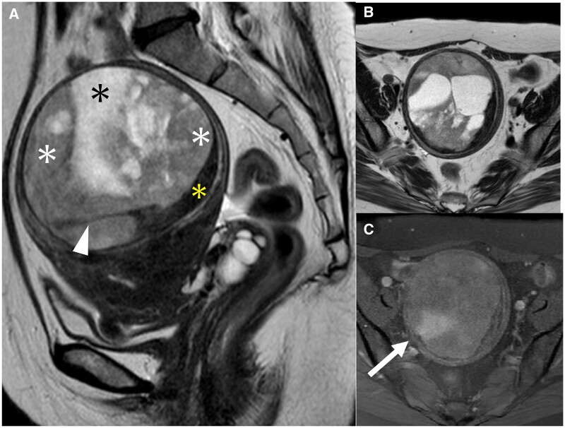
A low-grade endometrial stromal sarcoma (ESS) has a pattern of presenting as an intramyometrial mass and is often misdiagnosed as cellular leiomyoma or degenerative uterine leiomyoma. A low-grade ESS is a malignant tumour that requires total hysterectomy with bilateral salpingo-oophorectomy; while a leiomyoma is a benign tumour and could be acceptable for enucleation. As the treatment strategies differ between a low-grade ESS and leiomyoma, radiologists should be familiar with the characteristic MRI findings of a low-grade ESS. A 51-year-old woman with abnormal uterine bleeding had been observed for 2 years at a previous hospital for a uterine leiomyoma based on MRI findings. A contrast-enhanced MRI demonstrated an intramyometrial mass composed of three components with the hypointense rim on T2-weighted images (T2WI): the first component was a homogeneous solid structure with mild hyperintensity on T2WI with a low apparent diffusion coefficient value; the second component was cystic; the third component was a structure of low signal intensity on T2WI similar to the muscle. Although a degenerative uterine leiomyoma was a differential diagnosis, these MRI findings were suggestive of a low-grade ESS. A total abdominal hysterectomy, bilateral salpingo-oophorectomy, pelvic lymphadenectomy, and partial omentectomy were performed. The pathological diagnosis was a low-grade ESS. In a low-grade ESS, there are three major patterns of MRI findings: one of these patterns is the less popular but clinically important intramyometrial mass pattern, which can be misdiagnosed as a leiomyoma, and this case conformed to this pattern.

 求助内容:
求助内容: 应助结果提醒方式:


