{"title":"Superior outcomes of pullout repairs for medial meniscus posterior root tears in partial tear compared to complete radial tear.","authors":"Masanori Tamura, Takayuki Furumatsu, Yusuke Yokoyama, Naohiro Higashihara, Koki Kawada, Toshifumi Ozaki","doi":"10.1186/s43019-023-00206-1","DOIUrl":null,"url":null,"abstract":"<p><strong>Purpose: </strong>To reveal the outcomes of partial medial meniscus posterior root tears following transtibial pullout repair compared with the outcomes of complete radial meniscus posterior root tears.</p><p><strong>Materials and methods: </strong>We retrospectively evaluated 15 consecutive patients (male/female, 5/10; average age, 64.4 years) who underwent transtibial pullout repair for partial medial meniscus posterior root tears and compared their results with those of 86 consecutive patients who underwent the same surgery for complete medial meniscus posterior root tears. All patients underwent second-look arthroscopy on average 1 year postoperatively, and a semi-quantitative meniscal healing score (anteroposterior width, stability, and synovial coverage, total 10 points) was evaluated. Medial meniscus extrusion was evaluated preoperatively and at second-look arthroscopy.</p><p><strong>Results: </strong>Postoperative clinical scores were not significantly different in the short term. However, second-look arthroscopy revealed a significant difference in repaired meniscal stability (partial tear; 3.3 points, complete tear; 2.3 points, p < 0.001) and total meniscal healing scores (partial tear; 8.3 points, complete tear; 7.1 points, p < 0.001). Medial meniscus extrusion progression was significantly different (partial tear; 0.4 mm, complete tear; 1.0 mm, p < 0.001).</p><p><strong>Conclusion: </strong>Partial medial meniscus posterior root tears showed better meniscal healing and less medial meniscus extrusion progression following pullout repair than complete medial meniscus posterior root tears.</p>","PeriodicalId":36317,"journal":{"name":"Knee Surgery and Related Research","volume":"36 1","pages":"8"},"PeriodicalIF":4.4000,"publicationDate":"2024-02-08","publicationTypes":"Journal Article","fieldsOfStudy":null,"isOpenAccess":false,"openAccessPdf":"https://www.ncbi.nlm.nih.gov/pmc/articles/PMC10854085/pdf/","citationCount":"0","resultStr":null,"platform":"Semanticscholar","paperid":null,"PeriodicalName":"Knee Surgery and Related Research","FirstCategoryId":"1085","ListUrlMain":"https://doi.org/10.1186/s43019-023-00206-1","RegionNum":0,"RegionCategory":null,"ArticlePicture":[],"TitleCN":null,"AbstractTextCN":null,"PMCID":null,"EPubDate":"","PubModel":"","JCR":"Q2","JCRName":"Medicine","Score":null,"Total":0}
引用次数: 0
Abstract
Purpose: To reveal the outcomes of partial medial meniscus posterior root tears following transtibial pullout repair compared with the outcomes of complete radial meniscus posterior root tears.
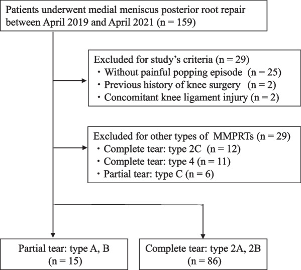
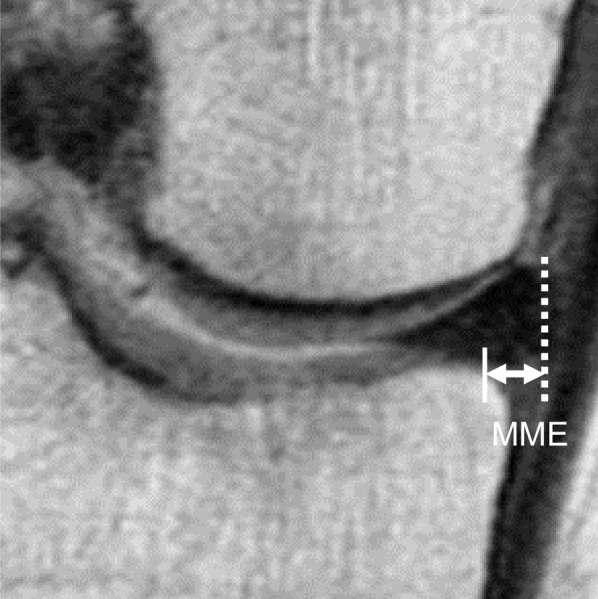
Materials and methods: We retrospectively evaluated 15 consecutive patients (male/female, 5/10; average age, 64.4 years) who underwent transtibial pullout repair for partial medial meniscus posterior root tears and compared their results with those of 86 consecutive patients who underwent the same surgery for complete medial meniscus posterior root tears. All patients underwent second-look arthroscopy on average 1 year postoperatively, and a semi-quantitative meniscal healing score (anteroposterior width, stability, and synovial coverage, total 10 points) was evaluated. Medial meniscus extrusion was evaluated preoperatively and at second-look arthroscopy.
Results: Postoperative clinical scores were not significantly different in the short term. However, second-look arthroscopy revealed a significant difference in repaired meniscal stability (partial tear; 3.3 points, complete tear; 2.3 points, p < 0.001) and total meniscal healing scores (partial tear; 8.3 points, complete tear; 7.1 points, p < 0.001). Medial meniscus extrusion progression was significantly different (partial tear; 0.4 mm, complete tear; 1.0 mm, p < 0.001).
Conclusion: Partial medial meniscus posterior root tears showed better meniscal healing and less medial meniscus extrusion progression following pullout repair than complete medial meniscus posterior root tears.

 求助内容:
求助内容: 应助结果提醒方式:


