{"title":"Fine Structure of the Gametes in Rhynchospio glandulosa (Annelida: Spionidae), a Hermaphrodite Brooding Larvae on the Parent’s Dorsum","authors":"V. I. Radashevsky, O. V. Yurchenko","doi":"10.1134/s1063074023080072","DOIUrl":null,"url":null,"abstract":"<h3 data-test=\"abstract-sub-heading\">Abstract</h3><p><i>Rhynchospio glandulosa</i> is a common polychaete living in silty tubes in soft sediments in temperate shallow waters in the Northwest Pacific. Worms are simultaneous hermaphrodites. Spermatogenesis occurs in the coelomic cavity. Spermatids are joined in 16-cell clusters. The spermatozoa have a dome-shaped acrosome 1.2 µm long, an elongated nucleus 2.9 µm long, midpiece 2.4 µm long with subspherical mitochondria, and a flagellum about 44 µm long. The acrosome is a complex structure with five internal parts. The nucleus is concave anteriorly and has a short fossa posteriorly which accommodates the basal body of the axoneme. The flagellum is strengthened by two circles of microtubules in addition to the central axoneme. Oogenesis is intraovarian. Developed oocytes are about 130 µm in diameter, with an envelope about 0.8 µm thick, consisting of three layers of extracellular matrix: the thickest basal layer penetrated by unbranched microvilli each about 0.4 µm long, a homogenous intermediate layer with the highest electron density, and the outer brush-like layer with numerous extensions each about 0.3 µm long. The oocytes are spawned to the parent’s dorsum where they are loosely held by flat branchiae and long dorsal capillaries. In this “hatchery” larvae develop until the four-segment stage, then leave the parent and continue development in sea water. The general morphology of long-headed spermatozoa and thin-envelope oocytes of <i>R. glandulosa</i> is similar to that of other brooding spionids, but the details of their gamete ultrastructure are different.</p>","PeriodicalId":49584,"journal":{"name":"Russian Journal of Marine Biology","volume":"277 2","pages":""},"PeriodicalIF":0.4000,"publicationDate":"2024-01-29","publicationTypes":"Journal Article","fieldsOfStudy":null,"isOpenAccess":false,"openAccessPdf":"","citationCount":"0","resultStr":null,"platform":"Semanticscholar","paperid":null,"PeriodicalName":"Russian Journal of Marine Biology","FirstCategoryId":"99","ListUrlMain":"https://doi.org/10.1134/s1063074023080072","RegionNum":4,"RegionCategory":"生物学","ArticlePicture":[],"TitleCN":null,"AbstractTextCN":null,"PMCID":null,"EPubDate":"","PubModel":"","JCR":"Q4","JCRName":"MARINE & FRESHWATER BIOLOGY","Score":null,"Total":0}
引用次数: 0
Abstract
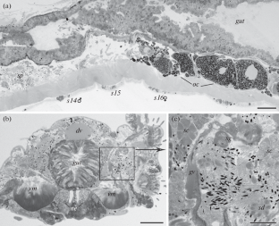
Rhynchospio glandulosa is a common polychaete living in silty tubes in soft sediments in temperate shallow waters in the Northwest Pacific. Worms are simultaneous hermaphrodites. Spermatogenesis occurs in the coelomic cavity. Spermatids are joined in 16-cell clusters. The spermatozoa have a dome-shaped acrosome 1.2 µm long, an elongated nucleus 2.9 µm long, midpiece 2.4 µm long with subspherical mitochondria, and a flagellum about 44 µm long. The acrosome is a complex structure with five internal parts. The nucleus is concave anteriorly and has a short fossa posteriorly which accommodates the basal body of the axoneme. The flagellum is strengthened by two circles of microtubules in addition to the central axoneme. Oogenesis is intraovarian. Developed oocytes are about 130 µm in diameter, with an envelope about 0.8 µm thick, consisting of three layers of extracellular matrix: the thickest basal layer penetrated by unbranched microvilli each about 0.4 µm long, a homogenous intermediate layer with the highest electron density, and the outer brush-like layer with numerous extensions each about 0.3 µm long. The oocytes are spawned to the parent’s dorsum where they are loosely held by flat branchiae and long dorsal capillaries. In this “hatchery” larvae develop until the four-segment stage, then leave the parent and continue development in sea water. The general morphology of long-headed spermatozoa and thin-envelope oocytes of R. glandulosa is similar to that of other brooding spionids, but the details of their gamete ultrastructure are different.
期刊介绍:
The Russian Journal of Marine Biology was founded in 1975 by Alexey V. Zhirmunsky, member of the Russian Academy of Sciences. The Russian Journal of Marine Biology covers a wide range of research and some applied aspects of marine biology as a synthetic science related to various fields of study on marine biota and environment. It presents fundamental research on biological processes at molecular, cellular, organismal, and populational levels in marine organisms. Consideration is given to marine objects as models in life sciences. The journal also publishes papers dedicated to events in Russian and international marine biological science and the history of biology.
 求助内容:
求助内容: 应助结果提醒方式:


