{"title":"Determining the safety margin of mandibular lingula in sagittal split ramus osteotomy","authors":"Gorkem Tekin, Nesrin Saruhan Kose, Mehmet Ugurlu, Omur Dereci, Yasin Caglar Kosar, Gunay Gojayeva, Gizem Caliskan","doi":"10.1007/s00276-023-03291-9","DOIUrl":null,"url":null,"abstract":"<h3 data-test=\"abstract-sub-heading\">Purpose</h3><p>The anatomical position of the lingula is clinically very important to prevent injuries during sagittal split ramus osteotomy. Our study aims to evaluate the localisation of the lingula by cone beam computed tomography (CBCT) and to compare the localisation of the lingula between malocclusion, gender, and lingula types.</p><h3 data-test=\"abstract-sub-heading\">Methods</h3><p>A retrospective study was conducted to evaluate the shape and location of the lingula using CBCT. A total of 250 CBCT images were included in this study. The lingula was classified as nodular, assimilated, truncated, or triangular type. Six defined distances from the top of the lingula were measured: anterior border of the ramus (L-A), posterior border of the ramus (L-P), internal oblique ridge (L-IOR), mandibular notch (L-N), and distal surface of the mandibular second molar (L-M2) and occlusal plane (L-OP). The measured distances were compared between gender, malocclusion, and lingula types.</p><h3 data-test=\"abstract-sub-heading\">Results</h3><p>The most common type of lingula was nodular (32.4%). The L-N, L-P, L-M2, and L-OP distances between genders were statistically higher in male patients than in female patients. The L-IOR, L-M2, and L-OP distances exhibited statistically significant differences found between malocclusions. No statistically significant difference was found when the distances of the lingula to the anatomical points were compared between the lingula types.</p><h3 data-test=\"abstract-sub-heading\">Conclusion</h3><p>These variations in positioning of the lingula depending on the dysmorphoses are developing towards a systematic 3D examination before any mandibular osteotomy to precisely visualize the position and shape of the lingula.</p>","PeriodicalId":49296,"journal":{"name":"Surgical and Radiologic Anatomy","volume":"48 1","pages":""},"PeriodicalIF":1.2000,"publicationDate":"2024-01-20","publicationTypes":"Journal Article","fieldsOfStudy":null,"isOpenAccess":false,"openAccessPdf":"","citationCount":"0","resultStr":null,"platform":"Semanticscholar","paperid":null,"PeriodicalName":"Surgical and Radiologic Anatomy","FirstCategoryId":"3","ListUrlMain":"https://doi.org/10.1007/s00276-023-03291-9","RegionNum":4,"RegionCategory":"医学","ArticlePicture":[],"TitleCN":null,"AbstractTextCN":null,"PMCID":null,"EPubDate":"","PubModel":"","JCR":"Q3","JCRName":"ANATOMY & MORPHOLOGY","Score":null,"Total":0}
引用次数: 0
Abstract
Purpose
The anatomical position of the lingula is clinically very important to prevent injuries during sagittal split ramus osteotomy. Our study aims to evaluate the localisation of the lingula by cone beam computed tomography (CBCT) and to compare the localisation of the lingula between malocclusion, gender, and lingula types.
Methods
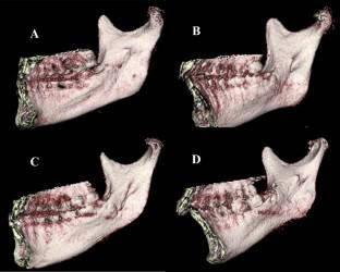
A retrospective study was conducted to evaluate the shape and location of the lingula using CBCT. A total of 250 CBCT images were included in this study. The lingula was classified as nodular, assimilated, truncated, or triangular type. Six defined distances from the top of the lingula were measured: anterior border of the ramus (L-A), posterior border of the ramus (L-P), internal oblique ridge (L-IOR), mandibular notch (L-N), and distal surface of the mandibular second molar (L-M2) and occlusal plane (L-OP). The measured distances were compared between gender, malocclusion, and lingula types.
Results
The most common type of lingula was nodular (32.4%). The L-N, L-P, L-M2, and L-OP distances between genders were statistically higher in male patients than in female patients. The L-IOR, L-M2, and L-OP distances exhibited statistically significant differences found between malocclusions. No statistically significant difference was found when the distances of the lingula to the anatomical points were compared between the lingula types.
Conclusion
These variations in positioning of the lingula depending on the dysmorphoses are developing towards a systematic 3D examination before any mandibular osteotomy to precisely visualize the position and shape of the lingula.
期刊介绍:
Anatomy is a morphological science which cannot fail to interest the clinician. The practical application of anatomical research to clinical problems necessitates special adaptation and selectivity in choosing from numerous international works. Although there is a tendency to believe that meaningful advances in anatomy are unlikely, constant revision is necessary. Surgical and Radiologic Anatomy, the first international journal of Clinical anatomy has been created in this spirit.
Its goal is to serve clinicians, regardless of speciality-physicians, surgeons, radiologists or other specialists-as an indispensable aid with which they can improve their knowledge of anatomy. Each issue includes: Original papers, review articles, articles on the anatomical bases of medical, surgical and radiological techniques, articles of normal radiologic anatomy, brief reviews of anatomical publications of clinical interest.
Particular attention is given to high quality illustrations, which are indispensable for a better understanding of anatomical problems.
Surgical and Radiologic Anatomy is a journal written by anatomists for clinicians with a special interest in anatomy.

 求助内容:
求助内容: 应助结果提醒方式:


