Light damage induces inflammatory factors in mouse retina and vitreous humor.
IF 1.4 3区 医学Q4 BIOCHEMISTRY & MOLECULAR BIOLOGY
Molecular VisionPub Date : 2023-10-15eCollection Date: 2023-01-01
Wei Liu, Xingfei Zhu, Xiangyu Ge, Yulin Chen, David Wan-Cheng Li, Lili Gong
{"title":"Light damage induces inflammatory factors in mouse retina and vitreous humor.","authors":"Wei Liu, Xingfei Zhu, Xiangyu Ge, Yulin Chen, David Wan-Cheng Li, Lili Gong","doi":"","DOIUrl":null,"url":null,"abstract":"<p><strong>Purpose: </strong>Increased inflammatory factor levels have been reported in the vitreous humor (VH) of diabetic retinopathy and neovascular age-related macular degeneration, ocular diseases generally associated with the formation of new retinal blood vessels and leakage. However, the levels of inflammatory mediators are less known in retinal degeneration without neovascularization. Human retinitis pigmentosa (RP) and animal models of light-induced retinal degeneration (LIRD) share several features, such as photoreceptor death and retinal inflammation. Here, we aimed to determine the levels of inflammatory factors in the VH of the LIRD mouse model.</p><p><strong>Methods: </strong>LIRD was induced by exposing BALB/c mice to white light (15,000 lx, 2 h), and the mice were recovered for 2 days before analysis (n = 50 mice). We assessed retinal morphology using optical coherence tomography and hematoxylin and eosin staining; retinal cell viability was determined using terminal deoxynucleotidyl transferase dUTP nick-end labeling, and retinal responses were measured based on electroretinogram signals. Total retinal RNAs were extracted and subjected to RNA sequencing analysis. VH samples from control (n = 4) and LIRD mice (n = 9) were assayed in triplicate for a panel of four inflammatory mediators using the Simple Plex Cartridge on an Ella System.</p><p><strong>Results: </strong>Retinal degeneration, photoreceptor death, infiltration of microglia/macrophages into the photoreceptor layer, and loss of a- and b-waves were obviously detected after LIRD. RNA sequencing revealed that light damage (LD) led to the significant upregulation of inflammatory factors in mouse retinas. In the VH, LD increased the total protein concentration. Dramatic induction of CCL2 (~3000 fold) and IL6 (~10 fold) was detected in VH in response to LD. Increased but not significant levels of TNFα and IL1β were also detected in light-exposed VH.</p><p><strong>Conclusions: </strong>Given that the LIRD model mimics RP pathogenesis in some aspects, these results suggest a causative link between retinal degeneration and VH inflammation in RP progression, and the increased CCL2 level in VH may reflect similar elevated CCL2 expression in the degenerative retina.</p>","PeriodicalId":18866,"journal":{"name":"Molecular Vision","volume":"29 ","pages":"180-187"},"PeriodicalIF":1.4000,"publicationDate":"2023-10-15","publicationTypes":"Journal Article","fieldsOfStudy":null,"isOpenAccess":false,"openAccessPdf":"https://www.ncbi.nlm.nih.gov/pmc/articles/PMC10784230/pdf/","citationCount":"0","resultStr":null,"platform":"Semanticscholar","paperid":null,"PeriodicalName":"Molecular Vision","FirstCategoryId":"3","ListUrlMain":"","RegionNum":3,"RegionCategory":"医学","ArticlePicture":[],"TitleCN":null,"AbstractTextCN":null,"PMCID":null,"EPubDate":"2023/1/1 0:00:00","PubModel":"eCollection","JCR":"Q4","JCRName":"BIOCHEMISTRY & MOLECULAR BIOLOGY","Score":null,"Total":0}
引用次数: 0
Abstract
Purpose: Increased inflammatory factor levels have been reported in the vitreous humor (VH) of diabetic retinopathy and neovascular age-related macular degeneration, ocular diseases generally associated with the formation of new retinal blood vessels and leakage. However, the levels of inflammatory mediators are less known in retinal degeneration without neovascularization. Human retinitis pigmentosa (RP) and animal models of light-induced retinal degeneration (LIRD) share several features, such as photoreceptor death and retinal inflammation. Here, we aimed to determine the levels of inflammatory factors in the VH of the LIRD mouse model.
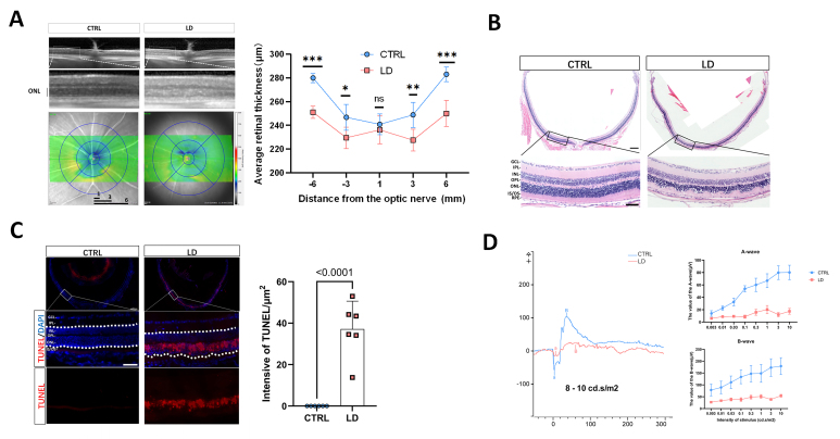
Methods: LIRD was induced by exposing BALB/c mice to white light (15,000 lx, 2 h), and the mice were recovered for 2 days before analysis (n = 50 mice). We assessed retinal morphology using optical coherence tomography and hematoxylin and eosin staining; retinal cell viability was determined using terminal deoxynucleotidyl transferase dUTP nick-end labeling, and retinal responses were measured based on electroretinogram signals. Total retinal RNAs were extracted and subjected to RNA sequencing analysis. VH samples from control (n = 4) and LIRD mice (n = 9) were assayed in triplicate for a panel of four inflammatory mediators using the Simple Plex Cartridge on an Ella System.
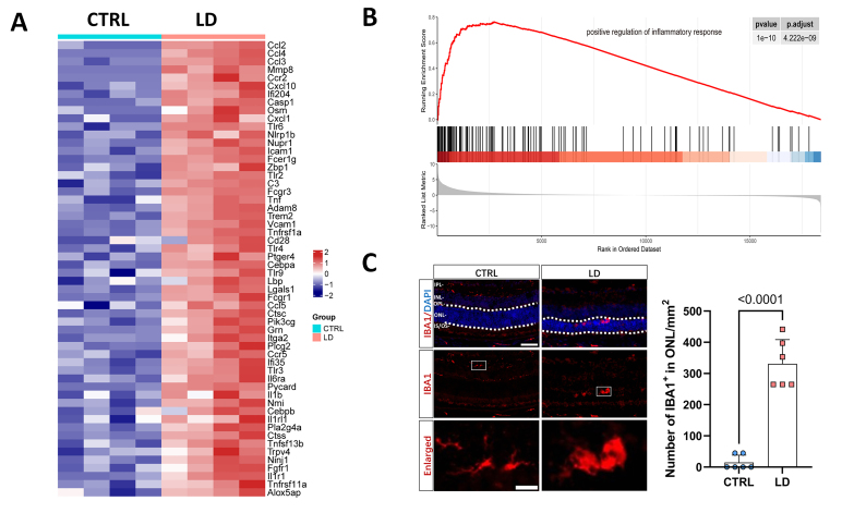
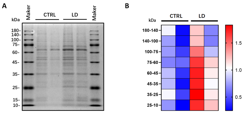
Results: Retinal degeneration, photoreceptor death, infiltration of microglia/macrophages into the photoreceptor layer, and loss of a- and b-waves were obviously detected after LIRD. RNA sequencing revealed that light damage (LD) led to the significant upregulation of inflammatory factors in mouse retinas. In the VH, LD increased the total protein concentration. Dramatic induction of CCL2 (~3000 fold) and IL6 (~10 fold) was detected in VH in response to LD. Increased but not significant levels of TNFα and IL1β were also detected in light-exposed VH.
Conclusions: Given that the LIRD model mimics RP pathogenesis in some aspects, these results suggest a causative link between retinal degeneration and VH inflammation in RP progression, and the increased CCL2 level in VH may reflect similar elevated CCL2 expression in the degenerative retina.
期刊介绍:
Molecular Vision is a peer-reviewed journal dedicated to the dissemination of research results in molecular biology, cell biology, and the genetics of the visual system (ocular and cortical).
Molecular Vision publishes articles presenting original research that has not previously been published and comprehensive articles reviewing the current status of a particular field or topic. Submissions to Molecular Vision are subjected to rigorous peer review. Molecular Vision does NOT publish preprints.
For authors, Molecular Vision provides a rapid means of communicating important results. Access to Molecular Vision is free and unrestricted, allowing the widest possible audience for your article. Digital publishing allows you to use color images freely (and without fees). Additionally, you may publish animations, sounds, or other supplementary information that clarifies or supports your article. Each of the authors of an article may also list an electronic mail address (which will be updated upon request) to give interested readers easy access to authors.
 求助内容:
求助内容: 应助结果提醒方式:


