Evaluation of the Algerbrush II rotating burr as a tool for inducing ocular surface failure in a mouse model.
IF 1.4 3区 医学Q4 BIOCHEMISTRY & MOLECULAR BIOLOGY
Molecular VisionPub Date : 2023-11-05eCollection Date: 2023-01-01
Athar Shadmani, Trent Jarin, Xiang Qi Meng, Yugendran Rajaendran, Salih Uzun, Albert Y Wu
{"title":"Evaluation of the Algerbrush II rotating burr as a tool for inducing ocular surface failure in a mouse model.","authors":"Athar Shadmani, Trent Jarin, Xiang Qi Meng, Yugendran Rajaendran, Salih Uzun, Albert Y Wu","doi":"","DOIUrl":null,"url":null,"abstract":"<p><strong>Purpose: </strong>The Algerbrush II has been widely used to induce corneal and limbal injuries in animal models. The extent of injury varies with the duration of exposure, pressure from the placement of the burr, and the size of the burr. However, no study has explored the correlation between the duration of exposure and the severity of injury in mouse model with corneal and limbal stem cell deficiency (LSCD) induced using the Algerbrush II. Therefore, this study aimed to evaluate the variations in the severity of corneal and limbal injury with different durations of the Algerbrush II application.</p><p><strong>Methods: </strong>The entire cornea and limbus of C57BL/6 mice were injured for 30-45 s, 60-75 s, 90-120 s, and 3-4 min. Photography and slit-lamp examination was performed on days 0, 2, 4, and 7, followed by hematoxylin & eosin, periodic acid-Schiff, and immunohistochemical staining. Statistical analysis was performed using one way ANOVA analysis.</p><p><strong>Results: </strong>A duration of 30-45 s of injury was found to be sufficient to induce superficial corneal and limbal epithelial debridement and re-epithelialization was completed in all eyes by day 7; however, clinical signs of LSCD were not observed in all mice. Increasing the exposure time to 90-120 s resulted in central 2+ corneal opacity with limbal and paracentral corneal neovascularization. All eyes injured for 3-4 min displayed clinical signs of LSCD, such as persistent epithelial defects on day 7 after the injury, central corneal neovascularization, and 2.2+ diffuse corneal opacity. Histological signs of LSCD, including goblet cell metaplasia and K13 expression on the corneal surface, were observed in all injured eyes.</p><p><strong>Conclusions: </strong>Our findings suggest that the duration of injury is an important factor influencing the severity of LSCD in a murine model of injury. A 1-mm rotating burr was found to be more effective for keratectomy and pigment release, whereas a 0.5-mm burr was more suitable for corneal epithelial debridement.</p>","PeriodicalId":18866,"journal":{"name":"Molecular Vision","volume":"29 ","pages":"256-265"},"PeriodicalIF":1.4000,"publicationDate":"2023-11-05","publicationTypes":"Journal Article","fieldsOfStudy":null,"isOpenAccess":false,"openAccessPdf":"https://www.ncbi.nlm.nih.gov/pmc/articles/PMC10784216/pdf/","citationCount":"0","resultStr":null,"platform":"Semanticscholar","paperid":null,"PeriodicalName":"Molecular Vision","FirstCategoryId":"3","ListUrlMain":"","RegionNum":3,"RegionCategory":"医学","ArticlePicture":[],"TitleCN":null,"AbstractTextCN":null,"PMCID":null,"EPubDate":"2023/1/1 0:00:00","PubModel":"eCollection","JCR":"Q4","JCRName":"BIOCHEMISTRY & MOLECULAR BIOLOGY","Score":null,"Total":0}
引用次数: 0
Abstract
Purpose: The Algerbrush II has been widely used to induce corneal and limbal injuries in animal models. The extent of injury varies with the duration of exposure, pressure from the placement of the burr, and the size of the burr. However, no study has explored the correlation between the duration of exposure and the severity of injury in mouse model with corneal and limbal stem cell deficiency (LSCD) induced using the Algerbrush II. Therefore, this study aimed to evaluate the variations in the severity of corneal and limbal injury with different durations of the Algerbrush II application.
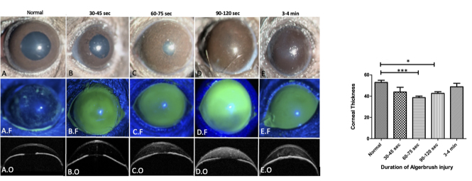
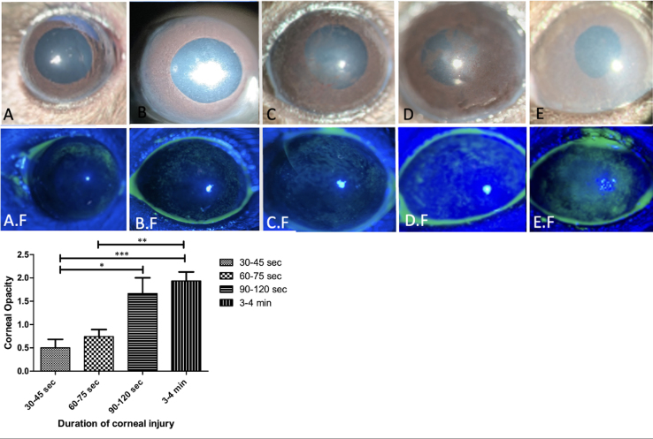
Methods: The entire cornea and limbus of C57BL/6 mice were injured for 30-45 s, 60-75 s, 90-120 s, and 3-4 min. Photography and slit-lamp examination was performed on days 0, 2, 4, and 7, followed by hematoxylin & eosin, periodic acid-Schiff, and immunohistochemical staining. Statistical analysis was performed using one way ANOVA analysis.
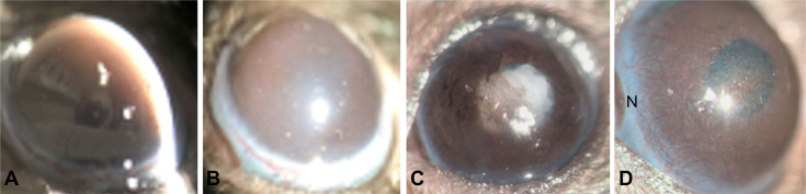
Results: A duration of 30-45 s of injury was found to be sufficient to induce superficial corneal and limbal epithelial debridement and re-epithelialization was completed in all eyes by day 7; however, clinical signs of LSCD were not observed in all mice. Increasing the exposure time to 90-120 s resulted in central 2+ corneal opacity with limbal and paracentral corneal neovascularization. All eyes injured for 3-4 min displayed clinical signs of LSCD, such as persistent epithelial defects on day 7 after the injury, central corneal neovascularization, and 2.2+ diffuse corneal opacity. Histological signs of LSCD, including goblet cell metaplasia and K13 expression on the corneal surface, were observed in all injured eyes.
Conclusions: Our findings suggest that the duration of injury is an important factor influencing the severity of LSCD in a murine model of injury. A 1-mm rotating burr was found to be more effective for keratectomy and pigment release, whereas a 0.5-mm burr was more suitable for corneal epithelial debridement.
期刊介绍:
Molecular Vision is a peer-reviewed journal dedicated to the dissemination of research results in molecular biology, cell biology, and the genetics of the visual system (ocular and cortical).
Molecular Vision publishes articles presenting original research that has not previously been published and comprehensive articles reviewing the current status of a particular field or topic. Submissions to Molecular Vision are subjected to rigorous peer review. Molecular Vision does NOT publish preprints.
For authors, Molecular Vision provides a rapid means of communicating important results. Access to Molecular Vision is free and unrestricted, allowing the widest possible audience for your article. Digital publishing allows you to use color images freely (and without fees). Additionally, you may publish animations, sounds, or other supplementary information that clarifies or supports your article. Each of the authors of an article may also list an electronic mail address (which will be updated upon request) to give interested readers easy access to authors.
 求助内容:
求助内容: 应助结果提醒方式:


