Daniel Berthe, Anna Kolb, Abdulrahman Rabi, Thorsten Sellerer, Villseveri Somerkivi, Georg Constantin Feuerriegel, Andreas Philipp Sauter, Felix Meurer, York Hämisch, Tuomas Pantsar, Henrik Lohman, Daniela Pfeiffer, Franz Pfeiffer
{"title":"Evaluation of Spectral X-Ray Imaging for Panoramic Dental Images Based on a Simulation Framework","authors":"Daniel Berthe, Anna Kolb, Abdulrahman Rabi, Thorsten Sellerer, Villseveri Somerkivi, Georg Constantin Feuerriegel, Andreas Philipp Sauter, Felix Meurer, York Hämisch, Tuomas Pantsar, Henrik Lohman, Daniela Pfeiffer, Franz Pfeiffer","doi":"10.1007/s10278-023-00940-8","DOIUrl":null,"url":null,"abstract":"<p>Modern photon counting detectors allow the calculation of virtual monoenergetic or material decomposed X-ray images but are not yet used for dental panoramic radiography systems. To assess the diagnostic potential and image quality of photon counting detectors in dental panoramic radiography, ethics approval from the local ethics committee was obtained for this retrospective study. Conventional CT scans of the head and neck region were segmented into bone and soft tissue. The resulting datasets were used to calculate panoramic equivalent thickness bone and soft tissue images by forward projection, using a geometry like that of conventional panoramic radiographic systems. The panoramic equivalent thickness images were utilized to generate synthetic conventional panoramic radiographs and panoramic virtual monoenergetic radiographs at various energies. The conventional, two virtual monoenergetic images at 40 keV and 60 keV, and material-separated bone and soft tissue panoramic equivalent thickness X-ray images simulated from 17 head CTs were evaluated in a reader study involving three experienced radiologists regarding their diagnostic value and image quality. Compared to conventional panoramic radiographs, the material-separated bone panoramic equivalent thickness image exhibits a higher image quality and diagnostic value in assessing the bone structure <span>\\(\\left(p<.001\\right)\\)</span> and details such as teeth or root canals <span>\\(\\left(p<.001\\right)\\)</span>. Panoramic virtual monoenergetic radiographs do not show a significant advantage over conventional panoramic radiographs. The conducted reader study shows the potential of spectral X-ray imaging for dental panoramic imaging to improve the diagnostic value and image quality.</p>","PeriodicalId":50214,"journal":{"name":"Journal of Digital Imaging","volume":"83 2 1","pages":""},"PeriodicalIF":2.9000,"publicationDate":"2024-01-12","publicationTypes":"Journal Article","fieldsOfStudy":null,"isOpenAccess":false,"openAccessPdf":"","citationCount":"0","resultStr":null,"platform":"Semanticscholar","paperid":null,"PeriodicalName":"Journal of Digital Imaging","FirstCategoryId":"5","ListUrlMain":"https://doi.org/10.1007/s10278-023-00940-8","RegionNum":2,"RegionCategory":"工程技术","ArticlePicture":[],"TitleCN":null,"AbstractTextCN":null,"PMCID":null,"EPubDate":"","PubModel":"","JCR":"Q2","JCRName":"RADIOLOGY, NUCLEAR MEDICINE & MEDICAL IMAGING","Score":null,"Total":0}
引用次数: 0
Abstract
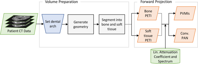
Modern photon counting detectors allow the calculation of virtual monoenergetic or material decomposed X-ray images but are not yet used for dental panoramic radiography systems. To assess the diagnostic potential and image quality of photon counting detectors in dental panoramic radiography, ethics approval from the local ethics committee was obtained for this retrospective study. Conventional CT scans of the head and neck region were segmented into bone and soft tissue. The resulting datasets were used to calculate panoramic equivalent thickness bone and soft tissue images by forward projection, using a geometry like that of conventional panoramic radiographic systems. The panoramic equivalent thickness images were utilized to generate synthetic conventional panoramic radiographs and panoramic virtual monoenergetic radiographs at various energies. The conventional, two virtual monoenergetic images at 40 keV and 60 keV, and material-separated bone and soft tissue panoramic equivalent thickness X-ray images simulated from 17 head CTs were evaluated in a reader study involving three experienced radiologists regarding their diagnostic value and image quality. Compared to conventional panoramic radiographs, the material-separated bone panoramic equivalent thickness image exhibits a higher image quality and diagnostic value in assessing the bone structure \(\left(p<.001\right)\) and details such as teeth or root canals \(\left(p<.001\right)\). Panoramic virtual monoenergetic radiographs do not show a significant advantage over conventional panoramic radiographs. The conducted reader study shows the potential of spectral X-ray imaging for dental panoramic imaging to improve the diagnostic value and image quality.
期刊介绍:
The Journal of Digital Imaging (JDI) is the official peer-reviewed journal of the Society for Imaging Informatics in Medicine (SIIM). JDI’s goal is to enhance the exchange of knowledge encompassed by the general topic of Imaging Informatics in Medicine such as research and practice in clinical, engineering, and information technologies and techniques in all medical imaging environments. JDI topics are of interest to researchers, developers, educators, physicians, and imaging informatics professionals.
Suggested Topics
PACS and component systems; imaging informatics for the enterprise; image-enabled electronic medical records; RIS and HIS; digital image acquisition; image processing; image data compression; 3D, visualization, and multimedia; speech recognition; computer-aided diagnosis; facilities design; imaging vocabularies and ontologies; Transforming the Radiological Interpretation Process (TRIP™); DICOM and other standards; workflow and process modeling and simulation; quality assurance; archive integrity and security; teleradiology; digital mammography; and radiological informatics education.
 求助内容:
求助内容: 应助结果提醒方式:


