Moritz Meyer-Jens , Kristin Wenzel , Karina Grube , Julia Rüdebusch , Elisabeth Krämer , Martin Bahls , Kilian Müller , Hannah Voß , Hartmut Schlüter , Stephan B. Felix , Lucie Carrier , Stephanie Könemann , Saskia Schlossarek
{"title":"Sacubitril/valsartan reduces proteasome activation and cardiomyocyte area in an experimental mouse model of hypertrophy","authors":"Moritz Meyer-Jens , Kristin Wenzel , Karina Grube , Julia Rüdebusch , Elisabeth Krämer , Martin Bahls , Kilian Müller , Hannah Voß , Hartmut Schlüter , Stephan B. Felix , Lucie Carrier , Stephanie Könemann , Saskia Schlossarek","doi":"10.1016/j.jmccpl.2023.100059","DOIUrl":null,"url":null,"abstract":"<div><p>Sacubitril/valsartan (Sac/Val) belongs to the group of angiotensin receptor–neprilysin inhibitors and has been used for the treatment of heart failure (HF) for several years. The mechanisms that mediate the beneficial effects of Sac/Val are not yet fully understood. In this study we investigated whether Sac/Val influences the two proteolytic systems, the ubiquitin-proteasome system (UPS) and the autophagy-lysosomal pathway (ALP), in a mouse model of pressure overload induced by transverse aortic constriction (TAC) and in human induced pluripotent stem cell-derived cardiomyocytes (hiPSC-CMs) treated with endothelin-1 (ET1) serving as a human cellular model of hypertrophy. TAC mice showed a continuous decline in cardiac function starting from day 14 after surgery. Administration of Sac/Val for 6 weeks counteracted the deterioration of cardiac function and attenuated hypertrophy and fibrosis in TAC mice. The expression of ALP key markers did not differ between the groups. Proteasome activity was higher in TAC mice and normalized by Sac/Val. In hiPSC-CMs, all treatments (Sac, Val or Sac/Val) normalized mean cell area. However, Sac alone or in combination with Val, but not Val alone prevented ET1-induced hypertrophic gene program and proteomic changes. In conclusion, Sac/Val normalized proteasome activity, improved cardiac function and reduced fibrosis and hypertrophy in TAC mice. Molecular analysis in hiPSC-CMs suggests that a major part of the beneficial effects of Sac/Val is derived from the Sac action rather than from Val.</p></div>","PeriodicalId":73835,"journal":{"name":"Journal of molecular and cellular cardiology plus","volume":"7 ","pages":"Article 100059"},"PeriodicalIF":0.0000,"publicationDate":"2024-01-07","publicationTypes":"Journal Article","fieldsOfStudy":null,"isOpenAccess":false,"openAccessPdf":"https://www.sciencedirect.com/science/article/pii/S2772976123000296/pdfft?md5=ac47fb87541aa8531ed0b59d80391286&pid=1-s2.0-S2772976123000296-main.pdf","citationCount":"0","resultStr":null,"platform":"Semanticscholar","paperid":null,"PeriodicalName":"Journal of molecular and cellular cardiology plus","FirstCategoryId":"1085","ListUrlMain":"https://www.sciencedirect.com/science/article/pii/S2772976123000296","RegionNum":0,"RegionCategory":null,"ArticlePicture":[],"TitleCN":null,"AbstractTextCN":null,"PMCID":null,"EPubDate":"","PubModel":"","JCR":"","JCRName":"","Score":null,"Total":0}
引用次数: 0
Abstract
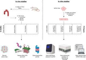
Sacubitril/valsartan (Sac/Val) belongs to the group of angiotensin receptor–neprilysin inhibitors and has been used for the treatment of heart failure (HF) for several years. The mechanisms that mediate the beneficial effects of Sac/Val are not yet fully understood. In this study we investigated whether Sac/Val influences the two proteolytic systems, the ubiquitin-proteasome system (UPS) and the autophagy-lysosomal pathway (ALP), in a mouse model of pressure overload induced by transverse aortic constriction (TAC) and in human induced pluripotent stem cell-derived cardiomyocytes (hiPSC-CMs) treated with endothelin-1 (ET1) serving as a human cellular model of hypertrophy. TAC mice showed a continuous decline in cardiac function starting from day 14 after surgery. Administration of Sac/Val for 6 weeks counteracted the deterioration of cardiac function and attenuated hypertrophy and fibrosis in TAC mice. The expression of ALP key markers did not differ between the groups. Proteasome activity was higher in TAC mice and normalized by Sac/Val. In hiPSC-CMs, all treatments (Sac, Val or Sac/Val) normalized mean cell area. However, Sac alone or in combination with Val, but not Val alone prevented ET1-induced hypertrophic gene program and proteomic changes. In conclusion, Sac/Val normalized proteasome activity, improved cardiac function and reduced fibrosis and hypertrophy in TAC mice. Molecular analysis in hiPSC-CMs suggests that a major part of the beneficial effects of Sac/Val is derived from the Sac action rather than from Val.

 求助内容:
求助内容: 应助结果提醒方式:


