Application of Machine Learning to Ultrasonography in Identifying Anatomical Landmarks for Cricothyroidotomy Among Female Adults: A Multi-center Prospective Observational Study
IF 3.8 2区 工程技术Q2 RADIOLOGY, NUCLEAR MEDICINE & MEDICAL IMAGING
{"title":"Application of Machine Learning to Ultrasonography in Identifying Anatomical Landmarks for Cricothyroidotomy Among Female Adults: A Multi-center Prospective Observational Study","authors":"Chih-Hung Wang, Jia-Da Li, Cheng-Yi Wu, Yu-Chen Wu, Joyce Tay, Meng-Che Wu, Ching-Hang Hsu, Yi-Kuan Liu, Chu-Song Chen, Chien-Hua Huang","doi":"10.1007/s10278-023-00929-3","DOIUrl":null,"url":null,"abstract":"<p>We aimed to develop machine learning (ML)-based algorithms to assist physicians in ultrasound-guided localization of cricoid cartilage (CC) and thyroid cartilage (TC) in cricothyroidotomy. Adult female volunteers were prospectively recruited from two hospitals between September and December, 2020. Ultrasonographic images were collected via a modified longitudinal technique. You Only Look Once (YOLOv5s), Faster Regions with Convolutional Neural Network features (Faster R-CNN), and Single Shot Detector (SSD) were selected as the model architectures. A total of 488 women (mean age: 36.0 years) participated in the study, contributing to a total of 292,053 frames of ultrasonographic images. The derived ML-based algorithms demonstrated excellent discriminative performance for the presence of CC (area under the receiver operating characteristic curve [AUC]: YOLOv5s, 0.989, 95% confidence interval [CI]: 0.982–0.994; Faster R-CNN, 0.986, 95% CI: 0.980–0.991; SSD, 0.968, 95% CI: 0.956–0.977) and TC (AUC: YOLOv5s, 0.989, 95% CI: 0.977–0.997; Faster R-CNN, 0.981, 95% CI: 0.965–0.991; SSD, 0.982, 95% CI: 0.973–0.990). Furthermore, in the frames where the model could correctly indicate the presence of CC or TC, it also accurately localized CC (intersection-over-union: YOLOv5s, 0.753, 95% CI: 0.739–0.765; Faster R-CNN, 0.720, 95% CI: 0.709–0.732; SSD, 0.739, 95% CI: 0.726–0.751) or TC (intersection-over-union: YOLOv5s, 0.739, 95% CI: 0.722–0.755; Faster R-CNN, 0.709, 95% CI: 0.687–0.730; SSD, 0.713, 95% CI: 0.695–0.730). The ML-based algorithms could identify anatomical landmarks for cricothyroidotomy in adult females with favorable discriminative and localization performance. Further studies are warranted to transfer this algorithm to hand-held portable ultrasound devices for clinical use.</p>","PeriodicalId":50214,"journal":{"name":"Journal of Digital Imaging","volume":"8 1","pages":""},"PeriodicalIF":3.8000,"publicationDate":"2024-01-10","publicationTypes":"Journal Article","fieldsOfStudy":null,"isOpenAccess":false,"openAccessPdf":"","citationCount":"0","resultStr":null,"platform":"Semanticscholar","paperid":null,"PeriodicalName":"Journal of Digital Imaging","FirstCategoryId":"5","ListUrlMain":"https://doi.org/10.1007/s10278-023-00929-3","RegionNum":2,"RegionCategory":"工程技术","ArticlePicture":[],"TitleCN":null,"AbstractTextCN":null,"PMCID":null,"EPubDate":"","PubModel":"","JCR":"Q2","JCRName":"RADIOLOGY, NUCLEAR MEDICINE & MEDICAL IMAGING","Score":null,"Total":0}
引用次数: 0
Abstract
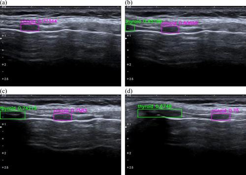
We aimed to develop machine learning (ML)-based algorithms to assist physicians in ultrasound-guided localization of cricoid cartilage (CC) and thyroid cartilage (TC) in cricothyroidotomy. Adult female volunteers were prospectively recruited from two hospitals between September and December, 2020. Ultrasonographic images were collected via a modified longitudinal technique. You Only Look Once (YOLOv5s), Faster Regions with Convolutional Neural Network features (Faster R-CNN), and Single Shot Detector (SSD) were selected as the model architectures. A total of 488 women (mean age: 36.0 years) participated in the study, contributing to a total of 292,053 frames of ultrasonographic images. The derived ML-based algorithms demonstrated excellent discriminative performance for the presence of CC (area under the receiver operating characteristic curve [AUC]: YOLOv5s, 0.989, 95% confidence interval [CI]: 0.982–0.994; Faster R-CNN, 0.986, 95% CI: 0.980–0.991; SSD, 0.968, 95% CI: 0.956–0.977) and TC (AUC: YOLOv5s, 0.989, 95% CI: 0.977–0.997; Faster R-CNN, 0.981, 95% CI: 0.965–0.991; SSD, 0.982, 95% CI: 0.973–0.990). Furthermore, in the frames where the model could correctly indicate the presence of CC or TC, it also accurately localized CC (intersection-over-union: YOLOv5s, 0.753, 95% CI: 0.739–0.765; Faster R-CNN, 0.720, 95% CI: 0.709–0.732; SSD, 0.739, 95% CI: 0.726–0.751) or TC (intersection-over-union: YOLOv5s, 0.739, 95% CI: 0.722–0.755; Faster R-CNN, 0.709, 95% CI: 0.687–0.730; SSD, 0.713, 95% CI: 0.695–0.730). The ML-based algorithms could identify anatomical landmarks for cricothyroidotomy in adult females with favorable discriminative and localization performance. Further studies are warranted to transfer this algorithm to hand-held portable ultrasound devices for clinical use.
期刊介绍:
The Journal of Digital Imaging (JDI) is the official peer-reviewed journal of the Society for Imaging Informatics in Medicine (SIIM). JDI’s goal is to enhance the exchange of knowledge encompassed by the general topic of Imaging Informatics in Medicine such as research and practice in clinical, engineering, and information technologies and techniques in all medical imaging environments. JDI topics are of interest to researchers, developers, educators, physicians, and imaging informatics professionals.
Suggested Topics
PACS and component systems; imaging informatics for the enterprise; image-enabled electronic medical records; RIS and HIS; digital image acquisition; image processing; image data compression; 3D, visualization, and multimedia; speech recognition; computer-aided diagnosis; facilities design; imaging vocabularies and ontologies; Transforming the Radiological Interpretation Process (TRIP™); DICOM and other standards; workflow and process modeling and simulation; quality assurance; archive integrity and security; teleradiology; digital mammography; and radiological informatics education.
 求助内容:
求助内容: 应助结果提醒方式:


