Orhan Beger, Baran Can Alpergin, Murat Zaimoglu, Ozgur Orhan, Mustafa Cemil Kılınç, Sena Unal, Halit Anil Eray, Umit Eroglu
{"title":"Massa intermedia in adults: incidence, dimension, location and clinical importance","authors":"Orhan Beger, Baran Can Alpergin, Murat Zaimoglu, Ozgur Orhan, Mustafa Cemil Kılınç, Sena Unal, Halit Anil Eray, Umit Eroglu","doi":"10.1007/s00276-023-03274-w","DOIUrl":null,"url":null,"abstract":"<h3 data-test=\"abstract-sub-heading\">Purpose</h3><p>This retrospective magnetic resonance imaging investigation aimed to obtain information related to the anatomy of the massa intermedia (MI) in an adult population.</p><h3 data-test=\"abstract-sub-heading\">Methods</h3><p>The work conducted on MRI views of 1058 (539 males and 519 females) healthy adult samples aged with 48.93 ± 17.63 years. Initially, the presence or absence of MI was noted, and then if present, its numbers and location in the third ventricle were recorded. Its horizontal (HDMI) and vertical (VDMI) diameters were measured on MRI views, while the cross-sectional area (CSAMI) was calculated using its diameters.</p><h3 data-test=\"abstract-sub-heading\">Results</h3><p>MI was missing in 2.6% (27 cases) of 1058 adult samples. Six subjects (0.6%) had a double MI. HDMI, VDMI and CSAMI were measured as 4.83 ± 1.01 mm, 4.86 ± 0.98 mm, and 19.11 ± 7.23 mm<sup>2</sup>, respectively. MI size did not show a significant alteration from 19 up to 49 years, but then its size distinctly decreased between 50 and 60 years. After age 60, MI dimension did not display an important change. MI was settled in the antero-superior quadrant in 929 cases (90.63% of 1025 subjects), in the postero-superior quadrant in 22 cases (2.15%), in the antero-inferior quadrant in 32 cases (3.12%), in the postero-inferior quadrant in 8 cases (0.78%), and in the central part in 34 cases (3.32%).</p><h3 data-test=\"abstract-sub-heading\">Conclusions</h3><p>The size, position and incidence of MI were not affected by sex, and its position and incidence were not affected by adult age periods. In adults, MI size demonstrated a significant decrease in the middle age.</p>","PeriodicalId":49296,"journal":{"name":"Surgical and Radiologic Anatomy","volume":"14 2 1","pages":""},"PeriodicalIF":1.2000,"publicationDate":"2024-01-08","publicationTypes":"Journal Article","fieldsOfStudy":null,"isOpenAccess":false,"openAccessPdf":"","citationCount":"0","resultStr":null,"platform":"Semanticscholar","paperid":null,"PeriodicalName":"Surgical and Radiologic Anatomy","FirstCategoryId":"3","ListUrlMain":"https://doi.org/10.1007/s00276-023-03274-w","RegionNum":4,"RegionCategory":"医学","ArticlePicture":[],"TitleCN":null,"AbstractTextCN":null,"PMCID":null,"EPubDate":"","PubModel":"","JCR":"Q3","JCRName":"ANATOMY & MORPHOLOGY","Score":null,"Total":0}
引用次数: 0
Abstract
Purpose
This retrospective magnetic resonance imaging investigation aimed to obtain information related to the anatomy of the massa intermedia (MI) in an adult population.
Methods
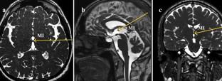
The work conducted on MRI views of 1058 (539 males and 519 females) healthy adult samples aged with 48.93 ± 17.63 years. Initially, the presence or absence of MI was noted, and then if present, its numbers and location in the third ventricle were recorded. Its horizontal (HDMI) and vertical (VDMI) diameters were measured on MRI views, while the cross-sectional area (CSAMI) was calculated using its diameters.
Results
MI was missing in 2.6% (27 cases) of 1058 adult samples. Six subjects (0.6%) had a double MI. HDMI, VDMI and CSAMI were measured as 4.83 ± 1.01 mm, 4.86 ± 0.98 mm, and 19.11 ± 7.23 mm2, respectively. MI size did not show a significant alteration from 19 up to 49 years, but then its size distinctly decreased between 50 and 60 years. After age 60, MI dimension did not display an important change. MI was settled in the antero-superior quadrant in 929 cases (90.63% of 1025 subjects), in the postero-superior quadrant in 22 cases (2.15%), in the antero-inferior quadrant in 32 cases (3.12%), in the postero-inferior quadrant in 8 cases (0.78%), and in the central part in 34 cases (3.32%).
Conclusions
The size, position and incidence of MI were not affected by sex, and its position and incidence were not affected by adult age periods. In adults, MI size demonstrated a significant decrease in the middle age.
期刊介绍:
Anatomy is a morphological science which cannot fail to interest the clinician. The practical application of anatomical research to clinical problems necessitates special adaptation and selectivity in choosing from numerous international works. Although there is a tendency to believe that meaningful advances in anatomy are unlikely, constant revision is necessary. Surgical and Radiologic Anatomy, the first international journal of Clinical anatomy has been created in this spirit.
Its goal is to serve clinicians, regardless of speciality-physicians, surgeons, radiologists or other specialists-as an indispensable aid with which they can improve their knowledge of anatomy. Each issue includes: Original papers, review articles, articles on the anatomical bases of medical, surgical and radiological techniques, articles of normal radiologic anatomy, brief reviews of anatomical publications of clinical interest.
Particular attention is given to high quality illustrations, which are indispensable for a better understanding of anatomical problems.
Surgical and Radiologic Anatomy is a journal written by anatomists for clinicians with a special interest in anatomy.

 求助内容:
求助内容: 应助结果提醒方式:


